Learn about First, Second, and Third degree AV heart blocks in this comprehensive ECG review. Each section will review the four different types of heart blocks and have a link to a review quiz afterwards.
Also, here is a lecture on heart blocks available to watch:
First-Degree AV Heart Block
Characteristics and Criteria of First-Degree Heart Block
This rhythm looks a lot like normal sinus rhythm, but it has a subtle secret. The PR intervals will be prolonged ( >0.20 seconds) regularly throughout the rhythm.

What you will see?
- Normal p-waves
- Normal atrial and ventricular rate (60-100 bpm) but can be slower
- Atrial and ventricular rhythm is regular
- Normal QRS <0.12 seconds
- Normal QT interval 0.36-0.44 seconds
- PR Interval >0.20 seconds regularly throughout the rhythm
Causes of First-Degree Heart Block
It can be normal for some patients. However, it can be present with a myocardial infarction (MI) or due to medications like calcium channel blockers, Digoxin, or beta blockers, which slow down AV node conduction.
Treatment for First-Degree Heart Block
Most patients are asymptomatic because this is usually detected randomly during a routine ECG reading.
If the patient has no symptoms, they will be monitored to make sure that it doesn’t progress to a more serious type of heart block or another abnormal rhythm. This is usually the least severe of all types of heart blocks.
The patient’s medications may have to be evaluated and adjusted, especially if they are taking a medication that can slow down AV node conduction like calcium channel blockers, beta blockers, or Digoxin.
In addition, if the patient has an extensive heart history a consult with a cardiologist may be needed for further evaluation.
Now test your knowledge on this material with this free First-Degree Heart Block Quiz.
ECG/EKG Study Guide and Workbook for Nursing Students
“ECG/EKG Interpretation Study Guide and Workbook by Nurse Sarah”. This book contain 100 pages of content featuring 26 ECG rhythm break downs, 51 ECG rhythm analysis practice problems, 100 comprehensive ECG practice questions, worksheets, chart summaries, and more.
You can get an eBook version here: “Nurse Sarah ECG Book” or a physical copy here: “ECG/EKG Interpretation Study Guide by Nurse Sarah“.
Second-Degree Type I AV Heart Block (Mobitz I or Wenckebach)
Characteristics and Criteria of Second-Degree Type I

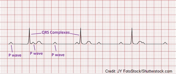
The key with this rhythm is that the PR interval follows a pattern where they gradually lengthen. Therefore, you will see that the p wave is getting further and further away from the QRS complex, and then all of the sudden there is a p wave without a QRS complex. Then this pattern will repeat itself again.
What you will see?
- Normal p-waves
- Atrial rate normal and rhythm regular
- Normal QRS complex <0.12 seconds
- Ventricular rate is slower than atrial rate and rhythm will be irregular (this is because QRS complexes are missing)
- Gradually lengthening or prolonged PR intervals until a QRS complex is dropped and then the cycle repeats

Causes of Second-Degree Type I
Myocardial infarction (especially during an active inferior wall MI due to active ischemia depriving the tissue of oxygen), medications that delay AV conduction like calcium channel blockers, Digoxin, beta blockers, rheumatic fever, and increased vagal tone
Treatment for Second-Degree Type I
Assess if having symptoms? If not, continue to monitor and an order may be given to consult with a cardiologist for further evaluation. Some medications may need to be stopped that slow AV conduction.
If patient is have signs and symptoms associated with a myocardial infarction (chest pain, dyspnea, sweating, clammy etc.) with this rhythm, get patient treatment FAST.
Therefore, if symptoms are presenting where cardiac output if falling (low blood pressure, weak pulse, mental status change, pale etc.) you need help so activate the emergency response team. Atropine or temporary pacing may be needed.
Now test your knowledge on this material by taking the free Second-Degree Type I Heart Block Quiz.
Second-Degree Type II (Mobitz II) Heart Block

Characteristics and Criteria for Second-Degree Type II Heart Block
The key with differentiating this rhythm from Second-Degree Type I is that with Second-Degree Type II the PR Intervals don’t progressively lengthen like with Type I, but stay constant, and then at some point a QRS complex is dropped. Furthermore, the PR interval can normal or prolonged.
What you will see?

- P-waves will be normal
- Atrial rate (60-100 bpm) and rhythm regular but some p waves won’t have a QRS complex after them. The QRS complex is MISSING.
- Ventricular rate will be slower than atrial rate (usually <60 bpm) and the rhythm irregular
- This is because some of the QRS complexes are missing.
- QRS complexes can be >0.12 seconds or <0.12 seconds depending on the blocked signal within the conduction system
- PR interval constant (the same throughout)…may be prolonged or normal
Causes of Second-Degree Type II Heart Block
Myocardial infarction with anterior wall involvement, advanced coronary artery disease, damage to the structures that make up the electrical conduction system, medications that slow AV conduction like calcium channel blockers, Digoxin, beta blockers etc.
Treatment for Second-Degree Type II Heart Block
This rhythm is worse than Second Degree Type I (Mobitz I/Wenckebach) and may progress to a third degree heart block.
With this rhythm the ventricular rate is on the slower side, which can affect cardiac output. Therefore, the patient is more likely to have symptoms with this rhythm. If no symptoms present, monitor closely and cardiology may need to be consulted for further evaluation. In addition, the assessment of the patient’s medications will be needed to ensure that they are not taking meds that slow down the AV conduction.
Symptomatic Treatment may include: temporary pacing and insertion of a permanent pacemaker
Now test your knowledge on this material by taking the Second-Degree Type II Heart Block Quiz.
Third-Degree (Complete) Heart Block ECG Review

Characteristics and Criteria of Third-Degree Heart Block
This is the worst type of heart block. The reason for this is because the electrical signal is not going from the atria to the ventricles. And this creates a major problem because the atria and ventricles are no longer working together.
Remember normally the signal goes from the atria to ventricles, creating a p wave (when the atria contract) and then a QRS complex (when the ventricles contract), and this happens continuously….a p wave always with a QRS complex.
However, this is not the case with a Third-Degree Heart Block. The p-waves are doing their own thing, and the QRS complexes are doing their own thing. Due to this cardiac output will become compromised.
What you will see?
- Normal p-waves but not found with every QRS complex (independent from QRS complexes)
- Normal atrial rhythm and rate
- Regular ventricular rhythm
- Slower ventricular rate than the atrial rate (less QRS complexes than p waves)
- Ventricular rate can be 40 bpm or less of depending on what is firing for the ventricles
- QRS complex width varies depending on what structure is firing…it can >0.12 or <0.12 seconds
- QRS complexes not always found after every p wave (independent from p waves)
- Variable PR intervals because the p waves and QRS complexes are independent
Causes of Third-Degree Heart Block
Congenital, heart disease, medications (Digoxin toxicity), structural damage to the heart, myocardial infarction, heart valve problem
Treatment of Third-Degree Heart Block
The patient is usually going to have symptoms that reflect impaired cardiac output. This symptoms can include hypotension, weak pulse, chest pain, pale, clammy etc.
Therefore, as the nurse you want to get help for your patient by activating the emergency response team. Atropine IV can be given to improve cardiac output. In addition, a temporary pacemaker, and then a permanent pacemaker can be implanted to prevent further problems.
Now test your knowledge on this material with the free Third-Degree (Complete) Heart Block Quiz.
Don’t forget about the Quizzes:
Second-Degree Type I Heart Block Quiz
Second-Degree Type II Heart Block Quiz
Third-Degree (Complete) Heart Block Quiz
References:
American Heart Association | Algorithms. (n.d.). Retrieved September 2, 2022, from https://cpr.heart.org/en/resuscitation-science/cpr-and-ecc-guidelines/algorithms#adult
Knabben V, Chhabra L, Slane M. Third-Degree Atrioventricular Block. [Updated 2022 May 22]. In: StatPearls [Internet]. Treasure Island (FL): StatPearls Publishing; 2022 Jan-. Available from: https://www.ncbi.nlm.nih.gov/books/NBK545199/
Mangi MA, Jones WM, Mansour MK, et al. Atrioventricular Block Second-Degree. [Updated 2022 May 22]. In: StatPearls [Internet]. Treasure Island (FL): StatPearls Publishing; 2022 Jan-. Available from: https://www.ncbi.nlm.nih.gov/books/NBK48235
Oldroyd SH, Quintanilla Rodriguez BS, Makaryus AN. First Degree Heart Block. [Updated 2022 May 1]. In: StatPearls [Internet]. Treasure Island (FL): StatPearls Publishing; 2022 Jan-. Available from: https://www.ncbi.nlm.nih.gov/b