This review will cover the differences between premature ventricular contractions (PVCs), premature atrial contractions (PACs), and premature junctional contractions (PJCs).
Don’t forget to check out all of our EKG interpretation reviews and to watch the lecture on PVCs vs. PACs vs. PJCs.
Premature Ventricular Contractions (PVCs)
Premature Ventricular Contractions (PVCs) are early contractions that originate in the ventricles, the bottom chambers of the heart. These contractions are typically caused by ventricular irritability. PVCs can occur suddenly and disrupt the normal electrical conduction process in the heart.
A typical PVC appears on an ECG as a wide and bizarre QRS complex, occurring usually within an underlying normal sinus rhythm.

For instance, above is an example of an underlying rhythm is a normal sinus rhythm with a premature ventricular contraction on the fourth beat and followed by this beat is a compensatory pause. This pause allows the heart to reset itself before the normal sinus rhythm resumes.
Key Characteristics of PVCs
When examining an ECG strip, several key characteristics of PVCs are evident:
- Wide and Bizarre QRS Complex: The QRS complex of a PVC is greater than 0.12 seconds and appears abnormal.
- Compensatory Pause: This pause follows the PVC, allowing the heart to prepare for the next beat.
- Irregular Rhythm: The rhythm becomes irregular due to the PVC, although the underlying rhythm outside of the PVC is typically regular.
- Absence of P-Wave: The P-wave is missing in PVCs, so the PR interval cannot be measured.
- Inaccurate QT Interval: Due to the wide QRS complex, the QT interval is difficult to measure accurately. An inverted T-wave may sometimes be seen after the PVC as well.
Types of PVCs
PVCs can vary in their presentation:

- Bigeminy: A normal beat followed by a PVC, occurring every other beat.
- Trigeminy: A pattern where a PVC occurs every third beat (normal beat, normal beat, PVC).
- Uniform vs. Multiform: PVCs can be uniform (similar in appearance) or multiform (different in appearance).
- Couplets: Two PVCs occurring consecutively.
When PVCs Become a Problem
Although PVCs are often benign, certain characteristics may indicate a need for further investigation:
- R-on-T Phenomenon: A PVC occurring on the T-wave of the preceding beat can increase the risk of ventricular tachycardia (VT) or ventricular fibrillation (VF), especially in patients with heart disease.
- Variability in Appearance: Multiform PVCs suggest they originate from different areas within the ventricles.
- Consecutive PVCs: Three or more consecutive PVCs, known as a run of ventricular tachycardia, warrant further evaluation.
- Pattern of PVCs: Patterns like bigeminy or trigeminy should be assessed for underlying conditions.
Causes of PVCs
Several factors can trigger PVCs, including:
- Caffeine Intake, Stress, Nicotine: Lifestyle factors that can contribute to PVCs
- Electrolyte Imbalances: Low potassium and magnesium levels.
- Myocardial Irritability: Conditions such as heart trauma or disease.
- Anemia, Thyroid Problems: Conditions like hyperthyroidism.
- Underlying Heart Conditions: Myocardial infarction, coronary artery disease, heart failure, rhythm disorders like atrial fibrillation, and bundle branch blocks.
- Elevated Blood Pressure: Can also contribute to PVCs.
Treatment of PVCs
PVCs are often asymptomatic and do not require treatment. However, when PVCs become frequent or symptomatic, treatment may be necessary. Symptoms of frequent PVCs include dizziness, syncope, palpitations, and a drop in blood pressure due to reduced cardiac output. To address these issues, treatment may involve:
- Managing Underlying Heart Conditions: Addressing the root cause of PVCs.
- Medications: Anti-arrhythmics like flecainide, beta-blockers, or calcium channel blockers can help reduce PVC frequency.
- Cardiac Ablation: If medications are ineffective, a cardiac ablation may be considered to eliminate the source of the PVCs.
Premature Atrial Contractions
Premature atrial contractions (PACs) are early heartbeats that originate from a focal point in the atria, rather than the sinoatrial (SA) node. These early beats cause the atria to contract prematurely. PACs are sometimes referred to as “PACs,” and they can be seen in a variety of rhythm patterns.
Identifying PACs
In a rhythm strip, PACs often appear as early P waves that look different from the regular, underlying P waves. Initially, there may be a normal sinus beat, followed by a premature beat. After these PACs, a brief pause may be noted before the rhythm resumes. This early P wave is usually different in shape or size compared to the normal P waves in the underlying rhythm.
PACs can be classified as either conducted or non-conducted. In conducted PACs, the early P wave is followed by a QRS complex, indicating that the electrical signal reached the ventricles and depolarized them. In non-conducted PACs, the P wave is not followed by a QRS complex, suggesting that the electrical signal did not reach the ventricles, possibly due to a block.
Characteristics of PACs
When observing PACs, the following characteristics are important:

- Irregular rhythm: The rhythm becomes irregular due to the PACs, but the underlying rhythm typically remains regular.
- Early P waves: The P waves of the PAC will differ in shape and size from the regular P waves in the underlying rhythm.
- QRS complex: The QRS complex is usually normal (less than 0.12 seconds), but may be missing if the PAC is non-conducted.
- PR interval: The PR interval may vary due to the PAC, as the premature P wave alters the timing of the electrical signal.
- QT interval: The QT interval may vary but is often normal.
- T-wave variation: The T wave may vary due to changes in ventricular repolarization after the PAC.
Causes of PACs
PACs can be caused by a variety of factors, including:
- Atrial enlargement, particularly of the left atrium
- Tobacco use and regular stimulant use, such as caffeine
- Inflammation of the atrial tissue
- Electrolyte imbalances, especially low potassium and magnesium levels
- Stress can also contribute to the occurrence of PACs
Symptoms and Treatment of PACs
PACs are often asymptomatic, and many patients may not be aware that they are having them, especially if they are infrequent. However, when PACs are frequent, patients may experience symptoms like palpitations or a fluttering sensation in the chest.
PACs can occur in patterns such as bigeminy (every other beat) or trigeminy (every third beat). The rhythm shown earlier was atrial bigeminy, where the rhythm alternates between normal sinus beats and PACs.
If PACs become frequent or if abnormal patterns are noted, further investigation may be necessary to rule out underlying heart conditions. Close monitoring of the rhythm is essential, and in some cases, medications such as beta-blockers and calcium channel blockers may be prescribed.
In addition to medication, it is important to address modifiable risk factors to prevent PACs. These include:
- Smoking cessation
- Limiting alcohol intake
- Staying hydrated
- Avoiding caffeine
- Managing stress
- Maintaining a healthy diet to help prevent electrolyte disturbances like low potassium and magnesium levels.
Premature Junctional Contractions (PJCs)
Premature junctional contractions (PJCs), also called premature junctional complexes, are early heartbeats that originate from the AV junction instead of the SA node.
What Are Premature Junctional Contractions (PJCs)?
PJCs are early contractions that occur prematurely within the underlying heart rhythm. Unlike normal beats that start at the SA node, PJCs originate at the AV junction, which is why they appear early and sometimes cause abnormal P-wave patterns.
Key Points to Remember:
- Premature Beats – PJCs occur before the next expected normal beat.
- P-Wave Abnormalities – PJCs can have unusual P-wave presentations:
- Concealed P-wave: Hidden within the QRS complex.
- Before the QRS: Appears very close, with a PR interval <0.12 seconds.
- After the QRS: Rare, but possible.
- Inverted P-wave in leads aVF, II, and III when present.

PJCs vs. Junctional Escape Beats
It’s important to differentiate PJCs from junctional escape beats:
- PJCs: Premature, random beats with no pause beforehand.
- Junctional Escape Beats: Occur after a pause, compensating for a slow SA node to prevent cardiac standstill.
ECG Characteristics of PJCs
When analyzing PJCs, look for the following:
Rhythm
- Underlying Rhythm: Regular
- PJC: Irregular due to the premature beat
Rate
- Underlying Rhythm: Depends on the underlying rhythm
- PJC: Same as underlying, but occurs early
P-wave
- Underlying Rhythm: Normal
- PJC: Can be inverted, hidden, or appear before/after QRS
PR interval
- Underlying Rhythm: 0.12–0.20 seconds
- PJC: Often <0.12 seconds or unmeasurable
QRS complex
- Underlying Rhythm: <0.12 seconds
- PJC: Normal, occurs early
QT interval & T-wave
- Underlying Rhythm: Normal
- PJC: Normal
Example: Sinus rhythm with occasional PJCs:

Sinus beat → Sinus beat → PJC (inverted P-wave, PR <0.12 sec) → Sinus beats → PJC (P-wave hidden in QRS)
Causes of PJCs
PJCs result from increased automaticity of the AV junction which can be due to the following: Digoxin toxicity / drugs, excessive alcohol use, oxygen deprivation / hypoxia, electrolyte imbalance (K⁺, Ca²⁺, Mg²⁺), tobacco use, injury to AV node (surgery, infection, congenital defects), can occur naturally in some patients
Nursing Considerations and Treatment
PJCs are usually asymptomatic and harmless when infrequent. Problems arise when they occur frequently, potentially reducing cardiac output and causing:
- Chest pain
- Palpitations
- Fluttering sensations
- Hypotension (if severe)
Assess underlying causes:
- Review medications (e.g., digoxin)
- Check electrolytes and hydration
- Identify lifestyle factors (smoking, alcohol, caffeine)
Patient Education:
- Limit alcohol and caffeine
- Avoid tobacco
- Stay hydrated
- Monitor medications that affect electrolytes (like diuretics)
Notify healthcare provider if PJCs are frequent or symptomatic.
Check digoxin levels if applicable:
- Therapeutic: 0.5–2 ng/mL
- Toxicity: Levels above 2 ng/mL
- Antidote: Digifab/Digibind
Key Takeaways for Nursing Students
- PJCs are premature beats from the AV junction.
- P-wave abnormalities are a hallmark feature.
- They differ from junctional escape beats because they occur without a preceding pause.
- Common causes include digoxin toxicity, AV node injury, electrolyte imbalances, hypoxia, and lifestyle factors.
- Treatment focuses on addressing underlying causes and patient education.
ECG/EKG Study Guide and Workbook for Nursing Students
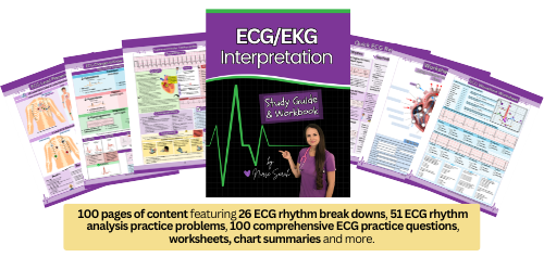
“ECG/EKG Interpretation Study Guide and Workbook by Nurse Sarah”. This book contain 100 pages of content featuring 26 ECG rhythm break downs, 51 ECG rhythm analysis practice problems, 100 comprehensive ECG practice questions, worksheets, chart summaries, and more.
You can get an eBook version here: “Nurse Sarah ECG Book” or a physical copy here: “ECG/EKG Interpretation Study Guide by Nurse Sarah“.
You may be interested in: ECG/EKG Identify Rhythms Practice Test
References:
American Heart Association. (n.d.). Premature contractions (PACs and PVCs). Retrieved June 26, 2024, from https://www.heart.org/en/health-topics/arrhythmia/about-arrhythmia/premature-contractions-pacs-and-pvcs
Amerman, E. C., & Irintcheva, V. (2016). Chapter 17 The Cardiovascular System I: The Heart. In Human Anatomy and Physiology (p. 636).
Farzam K, Richards JR. Premature Ventricular Contraction. [Updated 2023 Aug 8]. In: StatPearls [Internet]. Treasure Island (FL): StatPearls Publishing; 2024 Jan-. Available from: https://www.ncbi.nlm.nih.gov/books/NBK532991/
Hafeez Y, Grossman SA. Junctional Rhythm. [Updated 2023 Feb 5]. In: StatPearls [Internet]. Treasure Island (FL): StatPearls Publishing; 2024 Jan-. Available from: https://www.ncbi.nlm.nih.gov/books/NBK507715/
How the Heart Works | NHLBI, NIH. Retrieved 15 February 2022, from https://www.nhlbi.nih.gov/health-topics/how-heart-works
Thaler, M. S. (2010). Arrhythmias of Sinus Origin. In The Only EKG Book You’ll Ever Need (6th ed., pp. 110–111). essay, Lippincott, Williams, Wilkins.