In this anatomy and physiology lesson, I’m going to cover the only bone that makes up the anatomical arm, called the humerus, which is part of the appendicular skeleton.
Although the word “humerus” comes from a Latin word that means “shoulder, “ it is often referred to as the funny bone, because humerus (arm bone) and humorous (funny) are homophones (same sound, different spelling and meaning). In addition, when you hit your humerus, it can give a induce a funny “jarred” feeling, which is due to the nerves in that area.
The Anatomy of the Humerus
Most of you have probably heard the following expression: “Everything happens for a reason. “ When you look at the anatomy of the humerus, you’ll find that those knobs, grooves, and depressions are there for a reason.
So, with that thought in mind, let’s tackle the anatomy of the humerus bone.
Humerus Bone Anatomy
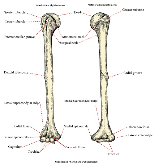
We’re going to be looking at a humerus from the right arm (pictured above), with the anterior (or front) view on the left, and the posterior (or back) view on the right.
- Head – Let’s start with the ball-shaped section at the proximal end (or top) of the humerus, which is called the head. It has a ball shape for a reason: it forms a ball-and-socket joint with the scapula (shoulder blade) at the glenoid cavity, which allows you to move your arm in various directions.
- Anatomical Neck – Immediately below the head, we have the anatomical neck of the humerus, which is a groove that provides a place for the shoulder’s joint capsule to attach.
- Greater and Lesser Tubercle – Next we have these small bumps called tubercles. Tubercles are small knobby structures on bones that allow for the attachment of muscles and ligaments. As the names suggest, the “greater” tubercle of the humerus is the “larger” knob structure located laterally. The “lesser” tubercle is the “smaller” knob, and it’s located medially. These tubercles allow for the attachment of muscles in your back, as well as the pectoralis major muscle.
- Intertubercular Groove – Anytime you have two hills side-by-side, you’re going to have a valley. And that’s what the intertubercular groove is – it’s a valley between the greater and lesser tubercles that extends down the shaft of the humerus. It’s easy to remember because its name is a dead giveaway: the prefix “inter” means “between,” and you only have two tubercles on the humerus, so this is found between those tubercles. This groove acts as a path for the long head of the biceps brachii.
- Surgical Neck – The part of the humerus that connects the larger proximal end (below the tubercles) to the skinnier shaft is called the surgical neck, not to be confused with the anatomical neck, which is the groove immediately below the head of the humerus. The surgical neck will often require surgical care, as this is one of the most common fracture points on the humerus!
- Radial Groove – On the posterior side (back) of the humerus, we have a groove called the radial groove. Why is that radial groove there? It houses the radial nerve.
- Deltoid Tuberosity – Next we have this odd, triangle-shaped projection coming off of the shaft of the humerus, which is called the deltoid tuberosity. Deltoid refers to the deltoid muscle, which attaches to this structure. Tuberosity is a fancy anatomy word that refers to a large prominence coming off a bone.
- Medial Supracondylar Ridge – Toward the distal end of the humerus’ shaft, we have a ridge that forms on each side, connecting to the epicondyles below. The ridge toward the midline of the body, which you can see in the posterior view of the right humerus above, is called the medial supracondylar ridge. Again, let the name help you. Medial is toward the midline of the body. The prefix supra means above, and condylar means knobby. So this is the ridge toward the middle of the body that is above the knobby structure at the end of the humerus. It allows for the attachment of muscles such as the brachialis and triceps brachii.
- Lateral Supracondylar Ridge – On the other side, we have the lateral supracondylar ridge, pictured here in the anterior view. Lateral means at the side or toward the side of the body. And this ridge allows for the attachment of various muscles such as the brachioradialis and triceps brachii.
- Coronoid Fossa – Next we have the coronoid fossa, which is near the middle of the humerus from the anterior view. Anytime you see the word fossa, it’s talking about a depression or crater-looking structure in the bone. Why is that depression there? When you flex your forearm, the apex of the coronoid process of the ulna (forearm bone) will fit into that depression on the humerus.
- Radial Fossa – The radial fossa is located laterally to the coronoid fossa. Guess what it does? It’s going to accomodate the head of the radius during forearm flexion.
- Olecranon Fossa – On the posterior side of the humerus, you’ll notice a huge depression called the olecranon fossa. Why is that depression there? When we extend our arm, that depression in the humerus is going to accommodate the olecranon process of the ulna, which is the bony projection that forms our elbow.
- Medial Epicondyle – Toward the distal end of the humerus, you can see how it fans out into a left and right knobby structure. The prefix “epi” tells us that this structure is upon or over, and condyle refers to the rounded protuberances known as the capitulum and trochlea. The medial epicondyle is toward the midline of the body, just over the trochlea.
- Lateral Epicondyle – The lateral epicondyle is located toward the side of the body, just over the capitulum. These structures allow for the attachment of various muscles of the forearm.
- Capitulum – The capitulum is a small, rounded protuberance (called a condyle) that is located laterally on the distal end of the humerus, which articulates (or forms a joint) with the head of the radius bone in the forearm.
- Trochlea – The trochlea is located medially and looks like a trophy on its side. The name trochlea refers to “spool” or “pulley,” and this part of the humerus articulates (or forms a joint) with the ulna bone of the forearm.
Free Quiz and More Anatomy Videos
Ready to test your knowledge? Take our free (and quick!) humerus anatomy quiz. Also, you might want to watch more of our anatomy and physiology lectures on YouTube, or check our anatomy and physiology notes.