This review explains the p wave on the ECG/EKG.
Before diving right into what a p wave is, we must go back and review the basic anatomy and physiology of the heart’s electrical conduction system.
Electrical Conduction System of the Heart:
It all starts in the:
SA Node (sinoatrial node): this node is located in the upper part the right atrium and is known as the pacemaker of the heart, causing the heart to beat at 60-100 bpm. When this node fires, it sends electrical impulses to the atria causing atrial depolarization of the cells in the right and left atrium (remember depolarization causes the atria to contract). Then the electrical signals go to the:
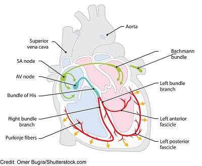
AV node (atrioventricular node): this node is found in the lower part of the right atrium just above the tricuspid valve and is known as the “gatekeeper”.
The AV node is known for causing a delay in electrical signaling so the atria can fully empty their blood into the ventricles. If there wasn’t a delay, the atria would not fully empty its blood into the ventricles and this would cause problems.
Then it’s time for the ventricles to be depolarized (hence contract). So the electrical signal goes down to the Bundle of His, then the bundle branches (right and left) and lastly the Purkinje fibers, which causes the ventricles to depolarize. Shortly after this process, repolarization occurs and this process repeats itself over and over again.
**If the electrical conduction system sends out electrical signals like it’s supposed to (with the electrical signaling starting out in the SA node and traveling down throughout the system) it will create the rhythm known as Normal Sinus Rhythm on the ECG strip. However, if there are problems within this system, dysrhythmias can occur like atrial fibrillation, atrial flutter, v-tach, etc.
ECG/EKG Study Guide and Workbook for Nursing Students
“ECG/EKG Interpretation Study Guide and Workbook by Nurse Sarah”. This book contain 100 pages of content featuring 26 ECG rhythm break downs, 51 ECG rhythm analysis practice problems, 100 comprehensive ECG practice questions, worksheets, chart summaries, and more.
You can get an eBook version here: “Nurse Sarah ECG Book” or a physical copy here: “ECG/EKG Interpretation Study Guide by Nurse Sarah“.
What’s the P Wave?
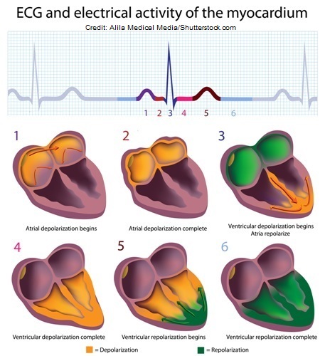
P wave: this represents atrial depolarization (leads to the atria contraction), which is created by the SA node. The atria receive blood and they must push it down to the ventricles by contracting. In other words, the p-wave is showing you that the atria are contracting.
How Should the P Wave Look?
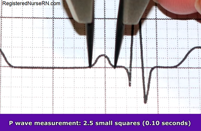
- One p-wave should be present and in front of every QRS complex, be upright and round, not flat
- Measurement should be less than 0.12 seconds (no more than 3 squares)
- Measuring from p wave to p wave details the atrial rhythm (is it regular or irregular)

- Counting the p waves within the strip details the atrial rate
- Atrial rate should be regular (60-100 bpm) to be a normal sinus rhythm and means it is originating in the SA node
You may be interested in more parts of the ECG Waveform.