Let’s take a look at the anatomy of a tooth, including its major structures and tissues.
But first, here’s a quick question that many people get wrong. Teeth are considered bones: true or false? The answer is…false. Although teeth and bones do have a similar appearance, they also have important differences. Therefore, anatomists do not classify teeth as bones.

Tooth Structure Anatomy
As you look at the anatomy of a tooth, you will see that it consists of three major parts: The crown, neck, and root.
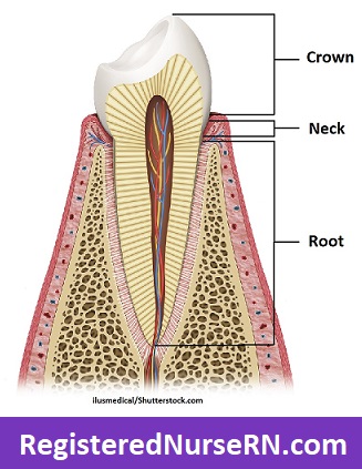
- The crown is the visible portion of the tooth above the neck and extending out of the gums. Just as you have a crown on your head, you can think of the tooth’s crown as the head (top) of the tooth. It is covered in a hard, protective layer called enamel, which covers the dentin and underlying pulp chamber and pulp.
- The neck of the tooth, also called the cementoenamel junction or cervical line, allows for the attachment of the gingivae (gums) to the teeth, and it represents the area where the crown’s enamel layer meets the cementum layer covering the tooth’s root. You can usually see the visible line of the neck when you look at an extracted tooth or a teeth with receded gums.
- The root is the larger portion of the tooth extending from the neck (cementoenamel junction) to the tooth’s apex, and it is secured to a socket called the alveolar process in the mandible (lower jaw bone) or maxilla (upper jaw bone). Some teeth, such as the incisors, have just one root, while others, such as the molars, can have two or three. Just as the crown is covered in a protective layer of enamel, the root is covered in its own protective layer called cementum, which covers the dentin and its underlying root canal and pulp.
Tooth Anatomy: Four Types of Tissues in the Tooth
The teeth may have three major parts, but these parts are comprised of four major tissue types: enamel, cementum, dentin, and pulp.

Tooth Enamel
Enamel, which makes up the hardened outer layer of the crown, does not contain living tissue such as vessels, nerves, or cells. Instead, is mostly comprised of a dense arrangement of minerals (around 95%) such as hydroxyapatite, which are formed into tiny prism rods that run in waves perpendicular to the tooth’s surface, though they can also run parallel near the tooth’s neck.

Although enamel is the hardest substance in the human body, it can dissolve after prolonged exposure to acid produced by dental plaque, which can eventually lead to the development of cavities (also called dental caries). Acidic foods can also temporarily soften enamel, which is why dental health professionals usually recommend waiting up to 30-60 minutes to brush your teeth after eating acidic foods.
Cementum
Cementum is the hardened bone-like tissue that covers the tooth’s root, and it’s formed by cells called cementoblasts. Cementum has a light yellow appearance and consists of a lower mineral concentration (around 45%) than both enamel and dentin, making it softer in comparison.
The periodontal ligament surrounds and attaches to the cementum of the tooth and anchors it to its socket in the alveolar process of the jawbone.
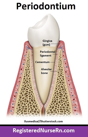
Cementum is also one of the four tissues that make up the periodontium, which surrounds and supports the teeth, the other three being the gingivae (gums), periodontal ligament, and the alveolar bone proper.
Dentin (or Dentine)
Dentin (also called dentine) is another hardened tissue of the tooth, and it is deep to both the crown’s enamel and the root’s cementum. It is formed and maintained by cells called odontoblasts. Dentin accounts for most of the tooth’s mass, and it contains a mineral content of around 70%, which makes it harder than cementum and even bone but softer than enamel.
Dentinal tubules run through the dentin in a parallel pattern, originating from the pulp cavity and ending just short of the enamel or cementum. These tubules transfer nutrients from the pulp to the dentin and contain an odontoblast process, which maintains the dentin.

Because these tubules originate at the pulp of the tooth, which contains the vessels and nerves, any damage to the enamel or gums can affect these tubules, which may result in tooth pain or sensitivity.
The border where the dentin meets the enamel is called the dentinoenamel junction (DEJ), and the border where the dentin meets the cementum is called the dentinocemental junction (DCJ).
Dental Pulp
Finally, the dental pulp is the soft tissue that is located within the pulp cavity of each tooth. The pulp cavity has two main parts: The narrow root canal of the root and the larger pulp chamber of the crown.
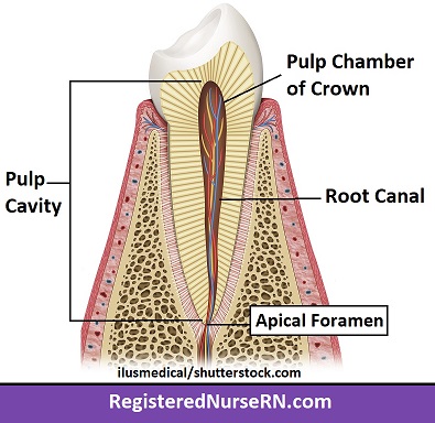
The root canal originates at a small hole in the apex of the root called the apical foramen, and it continues along the root(s) of the tooth. The root canal expands into a larger pulp chamber within the crown of the tooth.
The pulp’s tissue consists of odontoblasts, vessels, and nerves, which together provide the tooth’s nutrition, sensation, and immune defense, as well as the secretions that maintain the dentin.
Types of Teeth
Just as you have four main tissues that make up the teeth, you also have four types of teeth:
- incisors
- canines (cuspids)
- premolars (bicuspids)
- molars

I’ll talk more about the four types of teeth, as well as their functions and locations in my next lesson.
Free Quiz and More Anatomy Videos
Take a free comprehensive quiz on tooth anatomy to test your knowledge, or review our tooth anatomy video. In addition, you might want to watch our anatomy and physiology lectures on YouTube, or check our anatomy and physiology notes.