There are 3 types of muscle tissue in the human body: skeletal, smooth, and cardiac muscle. In this video and article compilation, I’m going to discuss all three types of muscle tissue.
Skeletal Muscle Tissue
Skeletal muscles most commonly attach to bones, and they help you move your body. Unlike the other two types of muscle tissue, skeletal muscles contract on a voluntary basis via the somatic nervous system, allowing you to move your body at will.
Skeletal muscles also serve important functions, such as supporting your posture, protecting delicate organs, and they even produce heat during contraction, which helps the body maintain a proper temperature.
Skeletal Muscle Structure
Each skeletal muscle is considered an organ, and it’s made up of connective tissue layers, muscle fibers, blood vessels, and nerves. Skeletal muscles attach to the bones through tendons or through a direct attachment.
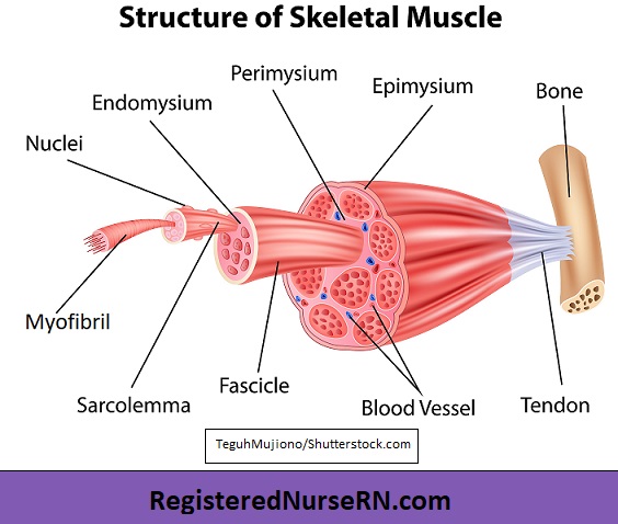
As you look at this muscle diagram, you’ll notice an outer layer of connective tissue called epimysium. The prefix “epi” means upon or over (epidermis is the layer upon your skin), and “mysium” comes from a Greek word that means “muscle.” Therefore, the epimysium is a layer of connective tissue that is over or upon the entire muscle organ.
Next, you’ll notice that the muscle fibers are bunched together into something called fascicles, which means “bundles.” These fascicles are surrounded by connective tissue called perimysium. “Peri” means “around,” and again, “mysium” refers to muscle. So the perimysium is around the fascicles that bundle up these muscle fibers.
Inside the fascicles, another connective tissue layer called the endomysium surrounds individual muscle cells. “Endo” means “within,” so that will help you remember that it surrounds the individual muscle fibers within the fascicle.
Muscle Fibers
Now let’s take a look at the individual muscle cells, which are called muscle fibers. These fibers are long and cylindrical, and they contain several nuclei. These muscle fibers are wrapped in a cell membrane called sarcolemma.
Inside each muscle fiber, there are tiny rods called myofibrils, which are surrounded by sarcoplasm. These myofibrils, also called fibrils, consist of repeating segments called sarcomeres, which are the tiny units responsible for skeletal muscle contraction.
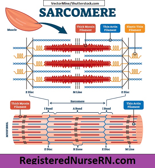
Sarcomere Structure
As we take a closer look at the structure of a sarcomere, you’ll notice these zigzag sections that mark the end point of each sarcomere. These are called Z discs or Z lines, and they allow for the attachment of the thin (actin) filaments, as well as an elastic protein called titin.
Each sarcomere contains thin (actin) filaments and thick (myosin) filaments. The thin (actin) filaments, represented below in blue, anchor to the Z disc.
These thick (myosin) filaments, represented below in red, attach to an elastic, springy protein called titin, which then attaches to the Z disc. The actin and myosin filaments engage during muscle contraction, which I’ll discuss in a moment. The “M lines” or “M bands” anchor the center of the myosin filaments, holding them together while also acting as a shock absorber.
Sarcomere Bands & Zones
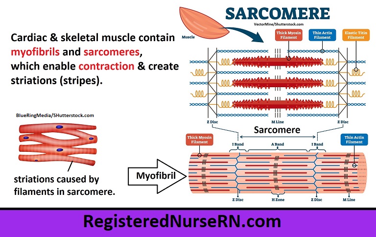
To help us understand the parts of the sarcomere, anatomists divide the sections into bands or zones. The arrangement of filaments within these bands accounts for the striated (striped) appearance of the skeletal muscle fibers. That’s an important characteristic about skeletal and cardiac muscle that you’ll want to remember: they both contain striations.
- A band: First, there is an “A band” on each sarcomere, which is a section that contains the entire length of a thick myosin filament, along with overlapping portions of the thin actin filaments. This section makes up the dark part of the striation pattern.
- I band: The “I band” is the section of the sarcomere that surrounds the Z disc and contains only thin (actin) filaments. This section makes up the lighter band in the striation pattern.
- H zone: The H zone is the section within the A band that consists of the thick myosin filaments and its embedded M lines. There are no thin filaments in this section of the sarcomere when it is relaxed.
- Z disc: And again, the Z disc is the zig zag portion that marks the end of each sarcomere and allows for the attachment of actin filaments and titin.
Skeletal Muscle Contraction
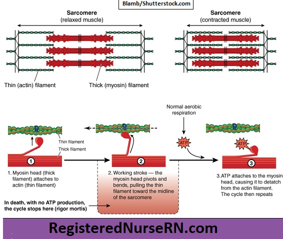
During muscle contraction, thick (myosin) filaments located within the sarcomere bend, and the knobby head part attaches to the thin actin filaments, sliding them toward the midline of the sarcomere. This sliding of filaments causes the sarcomere to shorten, or contract. As this takes place along all the sarcomeres within the myofibrils, the entire muscle fiber contracts, which ultimately causes the entire muscle organ to shorten or contract.
This is known as the sliding filament theory of muscle contraction.
Cardiac Muscle Tissue
Now let’s discuss another type of muscle tissue: cardiac muscle tissue. Of the three types of muscle tissue, cardiac muscle tissue is the one that’s near and dear to my heart because, well, it’s in my heart! In fact, that’s the only place you’ll find cardiac muscle tissue, and the very word “cardiac” literally means “relating to the heart.”
The cells that make up cardiac muscle are called cardiomyocytes (or cardiocytes), and together, they make up the myocardium, the muscle layer of the heart. This muscle layer causes the heart to contract in a wringing motion, which pumps blood throughout the body, supplying organs and tissues with oxygen and vital nutrients.
Cardiac muscle shares some similarities to skeletal muscle tissue, but there are also some key differences. First, let’s talk about the similarities.
Similarities of Cardiac and Skeletal Muscle Tissue
Cardiac Muscle Connective Tissue
Like skeletal muscle, cardiac muscle cells are surrounded and separated by a connective tissue layer called endomysium. Remember, “endo” means within, and “mysium” means muscle, so this is the connective tissue “within” or “closest to” the actual muscle cells or fibers.

Cardiac Muscle Contraction Mechanism
Cardiac muscle also contains myofibrils and sarcomeres, which not only enable contraction, but they also create the striations (striped pattern) that characterize both cardiac and skeletal muscle tissue. And that’s something you’ll want to remember: Both skeletal and cardiac muscle tissue have striations (or stripes)!
As I pointed out in my skeletal muscle video, sarcomeres contain actin and myosin filaments that slide during contraction, and the specific arrangement of these filaments into zones and bands creates that striped (or striated) appearance.
Differences of Cardiac vs Skeletal Muscle
Even though skeletal muscle and cardiac muscle have similarities, there are important differences between them.
Cardiac Muscle Contraction Control
Whereas skeletal muscle contracts on a voluntary basis (when you consciously want it to contract), cardiac muscle contracts on an INVOLUNTARY basis (without your conscious control) via the autonomic nervous system. That’s a good thing, because let’s be honest: if we had to consciously remember to make our heart pump, most of us would probably be dead by now.
Cardiac Muscle Shape
There is also a difference in shape between these muscle types. Skeletal muscle tissue develops into long, cylindrical fibers, but cardiac muscle tissue is formed into single cells that have an irregular branched appearance. These individual cells join to other cardiac cells via intercalated discs, which I’ll discuss in a moment.

Cardiac Muscle Nuclei
Another difference is that skeletal muscle contains multiple nuclei that are scattered around the peripheral portion of the muscle cells, whereas cardiac muscle contains only 1-2 nuclei, which are located near the center of the cell.
Intercalated Discs in Cardiac Muscle
Finally, an important characteristic of cardiac muscle cells that you’ll want to remember is that they are joined together by something called intercalated discs, which are absent in skeletal muscle tissue.
These intercalated discs form an interlocking zigzag connection between the individual cardiac muscle cells, and they consist of three types of cell junctions: desmosomes, fasciae adherens, and gap junctions.

- Desmosomes act as binders during contraction, supporting the filaments in adjoining cardiac cells to prevent separation.
- Fasciae adherens also work to connect and bind cardiac muscle cells by adhering to the actin filaments.
- Gap junctions are the tiny channels between adjoining cardiac cells that allow for the rapid passage of ions from once cell to the next, resulting in depolarization and contraction, which causes cardiac muscle cells to contract simultaneously. Specialized pacemaker cells in the heart connect to these gap junctions and work to control the heart rate.
Smooth Muscle Tissue
Finally, there is a third and final type of muscle tissue: smooth muscle tissue.
Smooth muscle is quite a bit different from the other two types of muscle tissue, but it also shares a few similarities. Here’s a quick rundown of the key concepts you’ll need to know about smooth muscle tissue.
Smooth Muscle Location
Whereas cardiac muscle is only located in the heart, and skeletal muscles mostly attach to bones, smooth muscle tissue is found throughout the body. To remember the main locations, I created a simple mnemonic to help you.
Remember the word “STOVE”:
- Skin (arrector pili muscles that cause goosebumps)
- Tracts found in the reproductive, respiratory, and urinary systems
- Organs that are hollow (such as the intestines, bladder, uterus, and stomach)
- Vessels (smooth muscle helps blood vessels constrict)
- Eyes (iris constriction/dilation, as well as the lens movement)
Smooth Muscle Shape and Orientation
Smooth muscle has a fusiform shape, which resembles a football or spindle. This is different from cardiac muscle tissue, which develops into an irregular branched pattern, or skeletal muscle tissue, which consists of fibers that are long and cylindrical. However, like skeletal and cardiac muscle, smooth muscle is also surrounded and separated by a connective tissue called endomysium.

Smooth Muscle Nucleus
Smooth muscle cells have only one nucleus, which is located in the central portion of the cell. In contrast, skeletal muscle tissue has multiple nuclei around the peripheral portion, whereas cardiac muscle usually has one or two nuclei centrally located.
Smooth Muscle Layers
Smooth muscle will often develop in layers within an organ to help it move in different directions. For example, in most of the digestive tract, smooth muscle cells are formed in two layers with different orientations, which work together to propel food down the digestive tract, a process known as peristalsis.
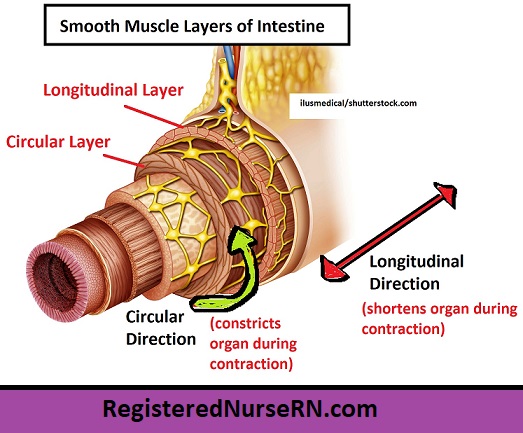
- Longitudinal layer – This word starts with “long,” and that will help you remember that these cells run along the length of the organ as the outermost smooth muscle layer, helping it become shorter during contraction.
- Circular layer – This layer is deep to the longitudinal layer and runs in a perpendicular direction to it, forming around the organ’s circumference, hence the word “circular.” This layer narrows (or constricts) the organ during contraction.
- Oblique layer – The stomach is unique in that it has a 3rd layer of smooth muscle, an oblique layer, which helps break down food before it reaches the intestines.
Smooth Muscle Control
Like cardiac muscle tissue, smooth muscle tissue is controlled involuntarily via the autonomic nervous system. This means that we do not consciously control it. Remember, skeletal muscle tissue is the only muscle tissue type that is voluntary (under our conscious control).
Smooth Muscle Structure
Smooth muscle tissue has a different structure compared to cardiac and skeletal muscle tissue. Smooth muscle does not contain sarcomeres, the organized contractile units that are found in cardiac and skeletal muscle tissue, nor does it contain the myofibrils, which are those rod-like structures made up of the repeating segments of sarcomeres.
Because smooth muscle lacks myofibrils and sarcomeres, it does not contain the striations (or striped pattern) that characterizes both skeletal and cardiac muscle tissue. And that’s an important point to remember for exams: smooth muscle is the only muscle tissue type that does not contain striations, and that’s why it’s called “smooth.”
However, smooth muscle tissue does consist of the same thin (actin) filaments and thick (myosin) filaments found in both skeletal and cardiac muscle tissue, which work to contract the muscle fiber via a sliding filament mechanism.
Smooth Muscle Anatomy
As you look at this diagram of a smooth muscle fiber, you’ll notice the single nucleus in the center. There is a net-like structure running throughout the muscle fiber. The little dots that connect the net structure are called dense bodies. Dense bodies attach to the sarcolemma, which is the smooth muscle cell’s outer sheath, and they work like the Z-disc in a sarcomere, allowing the thin filaments to attach to them.
The dense bodies also allow for the attachment of intermediate filaments such as desmin and vimentin, which run throughout the cell in a networked fashion, adding strength and stability to it.

Smooth Muscle Contraction
Smooth muscle contracts via a sliding filament mechanism, which is similar to that of skeletal and cardiac muscle. During contraction, calcium ions initiate a reaction that causes the phosphorylation of myosin, causing the heads on the myosin filaments to rise up and bind to the actin filaments, pulling them forward in the process.
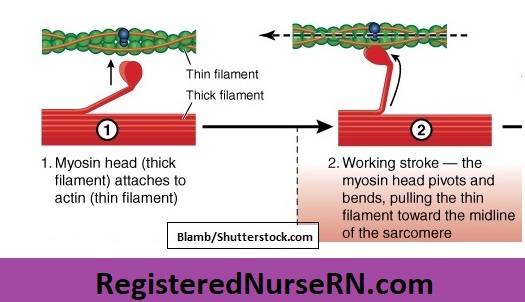
As the myosin filament heads slide the actin filaments forward, they also pull on the dense bodies to which the actin filaments attach, which then pulls on the network of intermediate filaments running throughout the cell. Thus, the entire smooth muscle fiber contracts, or shortens.

Single-unit vs Multi-unit Smooth Muscle
It’s important to note that there are actually two sub-types of smooth muscle tissue: single-unit and multi-unit smooth muscle. Single vs multi primarily refers to the number of nerve fibers required to activate the smooth muscle tissue.

- Single-unit smooth muscle, also called unitary smooth muscle, is innervated by only one (or very few) nerve fibers per bundle. There is no need for many nerve fibers, because one nerve fiber can contract the entire sheet of smooth muscle in unison due to the presence of gap junctions, which allow the electrical signal to spread rapidly to all of the adjacent smooth muscle cells. This would be analogous to a string of Christmas lights. A single plug could power all the lights in a strand because they are electrically coupled. Single-unit smooth muscle is found primarily in the hollow organs such as the intestines, which is why it is sometimes called visceral smooth muscle (viscera refers to organs or guts).
- Multi-unit smooth muscle, however, contains fewer (or no) gap junctions, so each cell requires its own electrical impulse (hence, there are “multiple” nerve fibers found in this type of smooth muscle). This would be analogous to separate lamps. Each lamp would require its own power source, hence, there would be “multiple” power sources needed due to the lack of electrical connection between the individual lamps. Multi-unit smooth muscle is found in the skin, eyes, blood vessels, and so on.
Free Quiz and More Anatomy Videos
Take a free muscle tissue types anatomy quiz to test your knowledge, or review our muscle tissue types video. In addition, you might want to watch our anatomy and physiology lectures on YouTube, or check our anatomy and physiology notes.