There are 3 types of muscle tissue in the human body: skeletal, smooth, and cardiac muscle. In this video and article, I’m going to discuss skeletal muscle tissue.
Skeletal Muscle Tissue
Skeletal muscles most commonly attach to bones, and they help you move your body. Unlike the other two types of muscle tissue, skeletal muscles contract on a voluntary basis via the somatic nervous system, allowing you to move your body at will.
Skeletal muscles also serve important functions, such as supporting your posture, protecting delicate organs, and they even produce heat during contraction, which helps the body maintain a proper temperature.
Skeletal Muscle Structure
Each skeletal muscle is considered an organ, and it’s made up of connective tissue layers, muscle fibers, blood vessels, and nerves. Skeletal muscles attach to the bones through tendons or through a direct attachment.
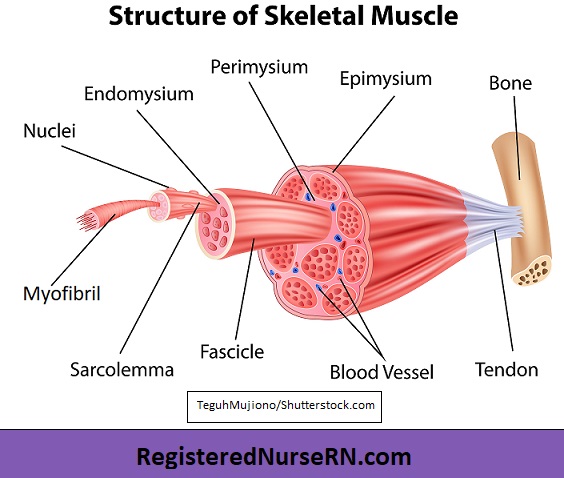
As you look at this muscle diagram, you’ll notice an outer layer of connective tissue called epimysium. The prefix “epi” means upon or over (epidermis is the layer upon your skin), and “mysium” comes from a Greek word that means “muscle.” Therefore, the epimysium is a layer of connective tissue that is over or upon the entire muscle organ.
Next, you’ll notice that the muscle fibers are bunched together into something called fascicles, which means “bundles.” These fascicles are surrounded by connective tissue called perimysium. “Peri” means “around,” and again, “mysium” refers to muscle. So the perimysium is around the fascicles that bundle up these muscle fibers.
Inside the fascicles, another connective tissue layer called the endomysium surrounds individual muscle cells. “Endo” means “within,” so that will help you remember that it surrounds the individual muscle fibers within the fascicle.
Muscle Fibers
Now let’s take a look at the individual muscle cells, which are called muscle fibers. These fibers are long and cylindrical, and they contain several nuclei. These muscle fibers are wrapped in a cell membrane called sarcolemma.
Inside each muscle fiber, there are tiny rods called myofibrils, which are surrounded by sarcoplasm. These myofibrils, also called fibrils, consist of repeating segments called sarcomeres, which are the tiny units responsible for skeletal muscle contraction.
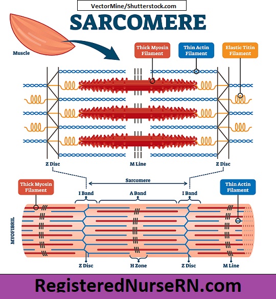
Sarcomere Structure
As we take a closer look at the structure of a sarcomere, you’ll notice these zigzag sections that mark the end point of each sarcomere. These are called Z discs or Z lines, and they allow for the attachment of the thin (actin) filaments, as well as an elastic protein called titin.
Each sarcomere contains thin (actin) filaments and thick (myosin) filaments. The thin (actin) filaments, represented below in blue, anchor to the Z disc.
These thick (myosin) filaments, represented below in red, attach to an elastic, springy protein called titin, which then attaches to the Z disc. The actin and myosin filaments engage during muscle contraction, which I’ll discuss in a moment. The “M lines” or “M bands” anchor the center of the myosin filaments, holding them together while also acting as a shock absorber.
Sarcomere Bands & Zones
To help us understand the parts of the sarcomere, anatomists divide the sections into bands or zones. The arrangement of filaments within these bands accounts for the striated (striped) appearance of the skeletal muscle fibers. That’s an important characteristic about skeletal and cardiac muscle that you’ll want to remember: they both contain striations.
- A band: First, there is an “A band” on each sarcomere, which is a section that contains the entire length of a thick myosin filament, along with overlapping portions of the thin actin filaments. This section makes up the dark part of the striation pattern.
- I band: The “I band” is the section of the sarcomere that surrounds the Z disc and contains only thin (actin) filaments. This section makes up the lighter band in the striation pattern.
- H zone: The H zone is the section within the A band that consists of the thick myosin filaments and its embedded M lines. There are no thin filaments in this section of the sarcomere when it is relaxed.
- Z disc: And again, the Z disc is the zig zag portion that marks the end of each sarcomere and allows for the attachment of actin filaments and titin.
Skeletal Muscle Contraction
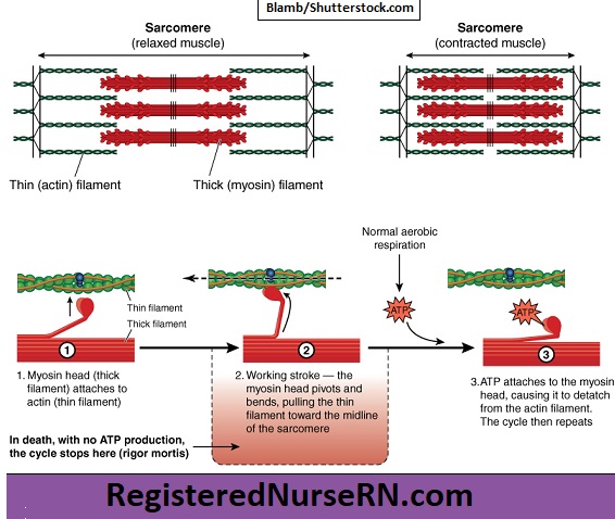
During muscle contraction, thick (myosin) filaments located within the sarcomere bend, and the knobby head part attaches to the thin actin filaments, sliding them toward the midline of the sarcomere. This sliding of filaments causes the sarcomere to shorten, or contract. As this takes place along all the sarcomeres within the myofibrils, the entire muscle fiber contracts, which ultimately causes the entire muscle organ to shorten or contract.
This is known as the sliding filament theory of muscle contraction.
Free Quiz and More Anatomy Videos
Take a free skeletal muscle anatomy quiz to test your knowledge, or review our skeletal muscle video. In addition, you might want to watch our anatomy and physiology lectures on YouTube, or check our anatomy and physiology notes.