In my last anatomy lesson, I discussed skeletal muscle tissue, which is one of the three types of muscle tissue found in the human body, along with cardiac and smooth muscle tissue. In this article and video, I’m going to discuss cardiac muscle tissue.
Of the three types of muscle tissue, cardiac muscle tissue is the one that’s near and dear to my heart because, well, it’s in my heart! In fact, that’s the only place you’ll find cardiac muscle tissue, and the very word “cardiac” literally means “relating to the heart.”
The cells that make up cardiac muscle are called cardiomyocytes (or cardiocytes), and together, they make up the myocardium, the muscle layer of the heart. This muscle layer causes the heart to contract in a wringing motion, which pumps blood throughout the body, supplying organs and tissues with oxygen and vital nutrients.
Cardiac muscle shares some similarities to skeletal muscle tissue, but there are also some key differences. First, let’s talk about the similarities.
Similarities of Cardiac and Skeletal Muscle Tissue
Cardiac Muscle Connective Tissue
Like skeletal muscle, cardiac muscle cells are surrounded and separated by a connective tissue layer called endomysium. Remember, “endo” means within, and “mysium” means muscle, so this is the connective tissue “within” or “closest to” the actual muscle cells or fibers.

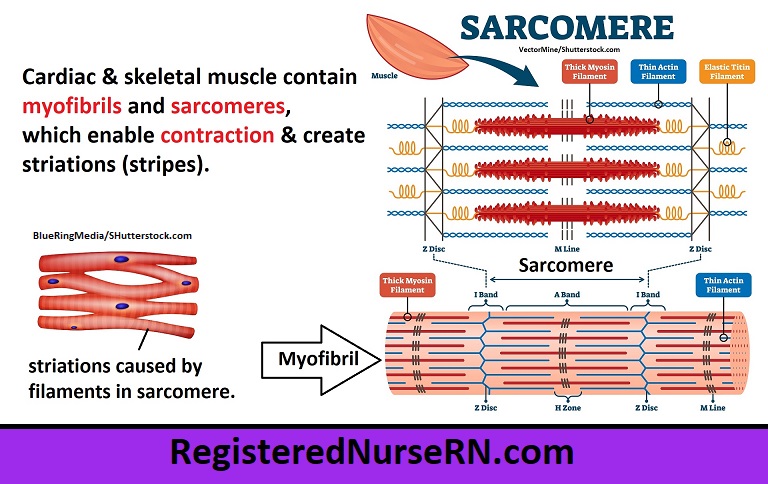
Cardiac Muscle Contraction Mechanism
Cardiac muscle also contains myofibrils and sarcomeres, which not only enable contraction, but they also create the striations (striped pattern) that characterize both cardiac and skeletal muscle tissue. And that’s something you’ll want to remember: Both skeletal and cardiac muscle tissue have striations (or stripes)!
As I pointed out in my skeletal muscle video, sarcomeres contain actin and myosin filaments that slide during contraction, and the specific arrangement of these filaments into zones and bands creates that striped (or striated) appearance.
Differences of Cardiac vs Skeletal Muscle
Even though skeletal muscle and cardiac muscle have similarities, there are important differences between them.
Cardiac Muscle Contraction Control
Whereas skeletal muscle contracts on a voluntary basis (when you consciously want it to contract), cardiac muscle contracts on an INVOLUNTARY basis (without your conscious control) via the autonomic nervous system. That’s a good thing, because let’s be honest: if we had to consciously remember to make our heart pump, most of us would probably be dead by now.
Cardiac Muscle Shape
There is also a difference in shape between these muscle types. Skeletal muscle tissue develops into long, cylindrical fibers, but cardiac muscle tissue is formed into single cells that have an irregular branched appearance. These individual cells join to other cardiac cells via intercalated discs, which I’ll discuss in a moment.

Cardiac Muscle Nuclei
Another difference is that skeletal muscle contains multiple nuclei that are scattered around the peripheral portion of the muscle cells, whereas cardiac muscle contains only 1-2 nuclei, which are located near the center of the cell.
Intercalated Discs in Cardiac Muscle
Finally, an important characteristic of cardiac muscle cells that you’ll want to remember is that they are joined together by something called intercalated discs, which are absent in skeletal muscle tissue.
These intercalated discs form an interlocking zigzag connection between the individual cardiac muscle cells, and they consist of three types of cell junctions: desmosomes, fasciae adherens, and gap junctions.

- Desmosomes act as binders during contraction, supporting the filaments in adjoining cardiac cells to prevent separation.
- Fasciae adherens also work to connect and bind cardiac muscle cells by adhering to the actin filaments.
- Gap junctions are the tiny channels between adjoining cardiac cells that allow for the rapid passage of ions from once cell to the next, resulting in depolarization and contraction, which causes cardiac muscle cells to contract simultaneously. Specialized pacemaker cells in the heart connect to these gap junctions and work to control the heart rate.
Free Quiz and More Anatomy Videos
Take a free cardiac muscle anatomy quiz to test your knowledge, or review our cardiac muscle video. In addition, you might want to watch our anatomy and physiology lectures on YouTube, or check our anatomy and physiology notes.