This review will discuss the heart’s anatomy, focusing on the chambers, great vessels, and valves.
The human heart is located in the mediastinum, a cavity between the right and left lungs. It is positioned slightly to the left of the center of the chest and situated behind the sternum.
People frequently imagine a heart as a literal heart shape located directly within the chest. However, the heart is more accurately shaped like an upside-down cone, with the base at the top and the pointed apex at the bottom, oriented slightly sideways.
The apex points more to the left side of the body, while the base is directed toward the right shoulder. In nursing, it’s important to be familiar with the base and apex, especially the apex, where the apical pulse is taken. This is the point of maximum impulse (PMI), indicating the strongest contraction.
Knowing the apex’s location is crucial because medications like digoxin require measuring the apical pulse beforehand. The apical pulse is typically located on the left side of the chest at the fifth intercostal space at the midclavicular line. Conversely, the base of the heart is located behind the sternum at the second intercostal space.
Chambers of the Heart
Let’s go inside the heart and examine its structure. In an anterior view of the heart, the left and right sides will appear reversed on your digital screen. The chambers include the right atrium at the top and the right ventricle below it on the right side. On the left side, the left atrium is the top chamber, with the left ventricle beneath it.

These chambers work together to pump blood from the right side to the left side of the heart. Blood on the right side is deoxygenated and travels through the pulmonary artery to the lungs, where it becomes oxygenated. It then returns to the left side of the heart via the pulmonary vein, entering the left atrium and passing through the left side to the aorta, from where it is pumped throughout the body.
For more on how blood flows through the heart.
Right Atrium and it’s Structures
Starting on the right side, the right atrium is the top chamber receiving deoxygenated blood from the superior vena cava, inferior vena cava, and coronary sinus. The walls of the right atrium are thinner than those of the left atrium due to lower pressure. The superior vena cava drains deoxygenated blood from the head, neck, upper chest, and upper extremities, while the inferior vena cava drains blood from the lower extremities, abdomen, and pelvic area. The coronary sinus opens into the right atrium, draining deoxygenated blood from the myocardium—the heart’s middle layer allowing it to contract. The fossa ovalis, found in the interatrial septum, was a passageway in the fetal heart allowing blood to bypass non-functioning lungs until birth. After birth, this area should close, leaving only an indentation. Failure to close can lead to a patent foramen ovale (PFO), potentially forming clots that cause strokes.
On the front inner surface of the right atrium, there are comb-like ridges referred to as pectinate muscles. The term “pectinate” comes from Latin, meaning “comb,” and as shown in the image above, they indeed resemble combs, much like the tool used to groom your hair.
These ridges have a purpose beyond aesthetics. They help the right atrium contract blood down into the right ventricle. These muscles are found in an area known as the auricle of the heart. The auricle is like a pouch-like extension that comes off of the atrium, and both the right and left atria have them.
You may wonder why this is termed an “auricle.” Remember, from medical terminology, that “auricle” refers to the outer ear. Use your imagination a little and look at these auricles; notice they sort of resemble two little flappy dog ears hanging off the side of each atrium. Not only do they help the atria contract blood down into the ventricles more efficiently, but they also act as extra storage space for blood. During increased physical activity, the heart can access that extra blood storage in those “flappy ears” to help maintain cardiac output.
Located near the pectinate muscles in the right atrium is an important smooth ridge called the crista terminalis. The crista terminalis plays a vital role in guiding how electrical impulses travel throughout the atria and from the SA node to the AV node, allowing the heart to contract rhythmically.
ECG/EKG Study Guide and Workbook for Nursing Students
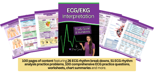
“ECG/EKG Interpretation Study Guide and Workbook by Nurse Sarah”. This book contain 100 pages of content featuring 26 ECG rhythm break downs, 51 ECG rhythm analysis practice problems, 100 comprehensive ECG practice questions, worksheets, chart summaries, and more.
You can get an eBook version here: “Nurse Sarah ECG Book” or a physical copy here: “ECG/EKG Interpretation Study Guide by Nurse Sarah“.
Right Ventricle and Nearby Structures
As we move down through the right atrium and open into the right ventricle, we encounter the tricuspid valve. The tricuspid valve is lined with endocardium, the inner layer of the heart, and has three strong cusps or flaps, hence its name. This valve is one of the two atrioventricular valves in the heart, the other being the mitral valve, also known as the bicuspid valve. These valves connect the atria and ventricles, which is why they are called atrioventricular.
The tricuspid valve allows blood to flow easily from the right atrium to the right ventricle while preventing backflow into the atria when the ventricles contract, ensuring a tight seal. The right ventricle is smaller and has thinner walls compared to the left ventricle.
It can have these attributes because it only needs to pump deoxygenated blood through pulmonary circulation, which typically has lower pressure. In contrast, the left ventricle is larger and thicker-walled because it must pump blood throughout the entire body, against a high-pressure systemic system. Important structures on the inner surface of the right ventricle aid its contraction, such as the trabeculae carneae. “Carneae” means “meaty,” and these are meaty ridges on the ventricle’s inner surface that provide support for proper contraction.
Within this area are also papillary muscles. “Papillary” refers to a protruding structure, similar to a nipple or cone. These muscles connect the tricuspid valve to the chordae tendineae, which are strong, string-like cords that create tension on the valve to keep it closed. They work with the papillary muscles to prevent valve collapse during ventricular contraction, ensuring proper blood flow direction by contracting when the ventricles contract.
Right after the right ventricle, you will find the pulmonic valve, also known as the pulmonary valve. This valve is one of the two semilunar valves in the heart. They are called semilunar because they are made up of three cusps that resemble crescent moons. These valves connect the ventricles to the great vessels and are lined with smooth endocardium.
These valves are not supported in the same way as atrioventricular valves, which use the chordae tendineae and papillary muscles. Instead, semilunar valves rely on pressure changes in the heart. For example, when the ventricles contract, the semilunar valves open, allowing blood to flow into the pulmonary artery or aorta. When the ventricles relax, pressure falls, causing the valves to shut, preventing backflow into the ventricles. The pulmonic valve connects the right ventricle and pulmonary artery, while the aortic valve connects the left ventricle and aorta. The semilunar valve’s name indicates which ventricle it connects to.
Taking a closer look at the pulmonic valve, we know its goal is to prevent backflow of blood into the right ventricle. It achieves this by depending on pressure changes within the right ventricle. When the right ventricle contracts, pressure increases inside, allowing the valve to open. Once open, blood is pushed through the valve into the pulmonary artery. As blood leaves, pressure drops, allowing the valve to close, preventing backflow into the right ventricle.
Now that blood has left the right ventricle, it enters the pulmonary artery. The pulmonary artery is sometimes referred to as the pulmonary trunk. It is unique because it carries deoxygenated blood away from the heart. While it is commonly said that arteries always carry oxygenated blood away from the heart, the pulmonary artery is an exception.
This artery starts as one and splits into two, the right and left, feeding into the right and left lungs. Its goal is to deliver deoxygenated blood to the lungs for oxygenation. The pulmonary artery walls are thinner than those of the aorta, another great vessel on the opposite side of the heart. This is to efficiently deliver blood to the lungs without high pressure, which could damage them over time. Once blood is oxygenated by the lungs, it returns to the left side of the heart via the pulmonary veins.
There are four pulmonary veins, with two sets coming from each lung and connecting to the left atrium. Like the pulmonary artery, the pulmonary veins are unique because they carry oxygenated blood back to the heart, specifically the left atrium.
Left Atrium and it’s Structures
The left atrium is mainly visible on the heart’s posterior side and is slightly smaller than the right atrium, with thicker walls. These thicker walls are necessary to contract against the high pressure on this side of the heart. The left atrium’s inner surface is smoother than the right atrium, with fewer pectinate muscles in its auricle. Oxygenated blood from the left atrium flows through the second atrioventricular valve, the mitral valve, also called the bicuspid valve. This valve has two cusps lined with endocardium.
To remember the location of each atrioventricular valve, think of the blood flow through the heart, starting on the right side, and remember the phrase “try before you buy.” The tricuspid valve comes first on the right side, and the bicuspid valve comes second on the left side.
From a nursing standpoint, familiarity with these heart valves is crucial, especially in cases of endocarditis. Endocarditis can be caused by IV drug use or severe infection entering the blood. As blood flows through the valves, germs can stick to them. I’ve assisted with transesophageal echocardiograms and seen young patients with large vegetations on their valves due to IV drug use, requiring heart valve replacement surgery and other procedures. Back to the mitral valve, it opens to allow blood flow from the left atrium to the left ventricle, preventing backflow into the left atrium. Like the tricuspid valve, it connects to the chordae tendineae and papillary muscles to stay in place during left ventricular contraction.
Left Ventricle and Nearby Structures
The left ventricle pumps oxygenated blood throughout the body, entering the aorta, another great vessel. Damage to this ventricle, such as from a myocardial infarction, can lead to heart failure. The interventricular septum separates the left ventricle from the right.
The left ventricle is larger and has thicker walls than the right, forming the heart’s apex. These attributes allow it to pump against the high-pressure systemic circulation. It contains trabeculae carneae, papillary muscles, and chordae tendineae, working similarly to the right ventricle.
Blood leaving the left ventricle passes through the second semilunar valve, the aortic valve, connecting the left ventricle and aorta. It functions like the pulmonary valve, relying on pressure changes for operation.
After passing through the aortic valve, blood enters the aorta, ascending and distributing oxygenated blood through branching arteries. One important branch includes the right and left coronary arteries, which wrap around the heart and supply the myocardium with fresh oxygenated blood.
This concludes a walkthrough of the heart’s anatomy. If you would like to learn more about the coronary arteries and heart layers, you can access the information via the provided links.
You may be interested in: heart anatomy lecture or heart anatomy quiz
References:
Amerman, E. C., & Irintcheva, V. (2016). Chapter 17 The Cardiovascular System I: The Heart. In Human Anatomy and Physiology (p. 636).
How the Heart Works | NHLBI, NIH. Retrieved 2 September 2022, from https://www.nhlbi.nih.gov/health-topics/how-heart-works
Marieb, E. N., & Mallatt, J. (2001). Chapter 18 The Heart. In Human Anatomy (Third, pp. 534–535).
Ogobuiro I, Wehrle CJ, Tuma F. Anatomy, Thorax, Heart Coronary Arteries. [Updated 2023 Jul 24]. In: StatPearls [Internet]. Treasure Island (FL): StatPearls Publishing; 2023 Jan-. Available from: https://www.ncbi.nlm.nih.gov/books/NBK534790/