This NCLEX review will discuss skin cancer.
Skin cancer occurs when cells in the skin turn cancerous. The types of skin cancer discussed in this lecture will be basal cell carcinoma, squamous cell carcinoma, actinic keratosis, and melanoma.
Don’t forget to take the skin cancer NCLEX questions quiz after reading this review.
Lecture on Skin Cancer
Skin Cancer NCLEX Review
What is skin cancer? It occurs when cells in the epidermis turn into cancerous cells.
There are different types of skin cancer, and the type of skin cancer is determined by the cell that has turned cancerous in the epidermis.
In this lecture we will review nonmelanoma and melanoma types of skin cancer.
- Nonmelanoma includes: basal cell carcinoma, squamous cell carcinoma….we will also discuss a precancerous form of skin cancer called actinic keratosis (solar keratosis)
- Melanoma
However, let’s quickly review the anatomy of the skin.
The layers of the skin include the epidermis, dermis, and hypodermis (subcutaneous fat).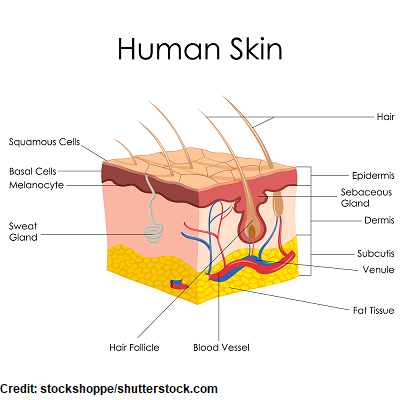
Some of the roles of the skin include regulating our body temperature, protecting us from germs, providing a tactile function to allow us to sense our environment, absorption (medication can be administered this way), and excretion of sweat etc.
Because we are learning about skin cancer in this review, we will be concentrating on the epidermis since this is where skin cancer originates. The epidermis is the top layer of the skin, and it’s divided into 5 layers. These 5 layers work together to help reconstruct our skin as we shed it.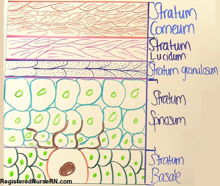
The 5 layers are (starting from top and going to the innermost layer):
- Stratum corneum
- Stratum lucidum
- Stratum granulosum
- Stratum spinosum
- Stratum basale
Let’s start with the bottom layer, stratum basale…also called the basal cell layer. It contains the basal cells, which work to create new skin cells. This is where basal cell carcinoma develops because these basal cells mutate into cancerous cells.
Also, located in the basal cell layer are melanocytes. These cells produce melanin (a dark brownish substance), which give us the color of our skin, hair, and eye. The neat thing about these cells are that when our skin is exposed to sunlight it causes the melanocytes to produce melanin, which will darken the skin to help protect it from the sun exposure….hence tanning the skin.
Melanoma occurs when the melanocytes become cancerous…hence why most lesions of melanoma are dark in color.
Then sitting on top of the basal cell layer is called the stratum spinosum (also called the squamous cell layer). A type of cells found in this layer is called keratinocytes. These cells produce keratin to make cells stronger so they stay together to provide a protective layer. This is where squamous cell carcinoma develops.
The other layers of the epidermis are the stratum granulosum, stratum lucidum, and stratum corneum, which is the very top layer you see when you look at your skin.
Types of Skin Cancer
*for exams be familiar with the descriptive words to describe each type
Nonmelanoma: these are types of skin cancer that do NOT originate from melanocytes so they’re not considered melanoma:
Basal Cell Carcinoma: most common form of skin cancer…appearance: “pearly” glossy, shiny, waxy, small raised bump with a depressed center and slightly elevated border.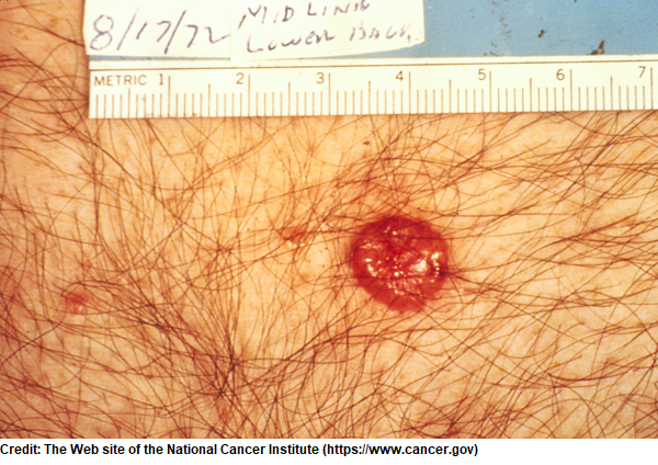
- It’s slow growing and metastasis (meaning the cancer spreads to other organs) is very rare….it originates from the basal cells in epidermis.
Squamous Cell Carcinoma: appearance: “crusty” hard-covering, scaly, pink or reddish and raised.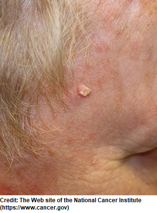
- It’s faster growing that basal cell and can metastasize…it originates from keratinocytes found in the squamous layer.
Actinic Keratosis (solar keratosis): appearance: scaly reddened patches 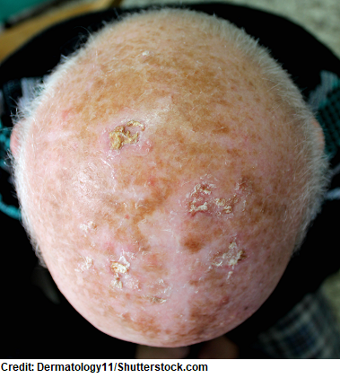
- It’s precancerous and can develop into squamous cell carcinoma, if not removed.
- It tends to affect older white adults and forms in areas exposed to the sun like the top of the head, face, arms etc.
Melanoma: appearance: use the ABCDE acronym to help detect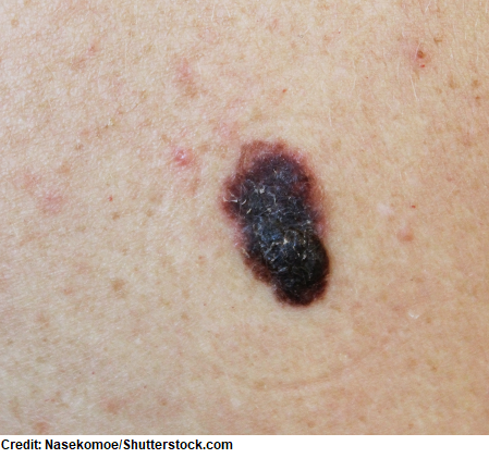
- asymmetrical (if a line is placed in the middle of the lesion it would NOT look the same)
- irregular borders
- dark colors (red, tan, black) or multiple
- diameter greater than 6mm
- evolving in shape/size/color
- It’s the most deadly form of skin cancer and has a very high risk of metastasizing (brain, lungs, bones etc.), if not detected early.
- It can form anywhere there are melanocytes (example: eyes in the middle layer called the uvea)
Risk Factors for Skin Cancer:
- Too much sun exposure (using tanning beds)
- Predisposition due to genetics (melanoma can be more common in certain families)
- Light skin, blonde, red hair, green or blue eyes
- Exposure to toxic chemicals
- History of frequent sunburns as a child
Nursing Interventions for Skin Cancer
We play a vital role in detection and education!
Asses the patient for any areas on the skin that doesn’t heal, itchy, or changes colors, and teach the patient to report this immediately to their doctor.
Perform a thorough skin assessment and identify possible cancerous lesions…follow the ABCDE assessment (teach the patient to do this monthly):

- Asymmetrical: if you draw a line in the middle of it, it should look the same on each side…abnormal would be that it doesn’t look the same (asymmetrical)
- Border are uneven
- Color: watch out for dark black or multiple colors
- Diameter: greater than 6mm
- Evolution: changes in size, shape, and color
Prevention (teach this to the patient):
- Avoid direct sun exposure between 10 am – 4 pm (sun rays are the strongest during these times)
- Wear long-sleeves, sun glasses, and a hat to avoid unnecessary sun exposure.
- Use a sunscreen that is broad-spectrum with a SPF of 15 or higher for exposed areas when outside.
- Avoid tanning beds and toxic chemical on the skin.
Treatment depends on the type of cancer and stage. However, the cancerous tissue will be removed (there are various ways to do this)….examples:
- cryosurgery (freezes off the cancerous tissue with liquid nitrogen)
- radiation (used for difficult to treat areas and will kill the cancerous cells)
- chemotherapy (kill cancerous cells)…topical: if hasn’t spread or systematic chemo if it has spread
- electrodessication and curettage (EDC): scraps and removes the abnormal tissue and an electrical current is used to prevent bleeding and to kill the cancerous cells…this is repeated a few times…Nursing care for surgical site: cover with petroleum jelly and nonstick bandage
References
Layers of the Skin | SEER Training. Retrieved from https://training.seer.cancer.gov/melanoma/anatomy/layers.html
Skin Cancer. Retrieved from https://www.cancer.gov/types/skin
What Can I Do to Reduce My Risk of Skin Cancer? | CDC. Retrieved from https://www.cdc.gov/cancer/skin/basic_info/prevention.htm