In this anatomy lesson, I’m going to cover the sacrum, a triangular bone located near the inferior portion of the vertebral column.
Sacrum Etymology
The word “sacrum” sounds a lot like the word “sacred,” and there’s a reason for that. “Sacrum” comes from an ancient Greek word that means “sacred bone” (hieron osteon), and they believed that this bone housed the soul.
Sacrum Classification
Anatomists classify the sacrum as an irregular bone, and it is located within the axial skeleton. It articulates superiorly (above) with the fifth lumbar vertebra, laterally (at the sides) with the bones of the hip (os coxae), and inferiorly (below) with the coccyx bone.
The sacrum bone begins as five separate vertebrae, which anatomists label as S1-S5, but they later fuse into one sacrum bone during early adulthood. In fact, when looking the sacrum’s anterior surface, you’ll notice four horizontal lines called transverse lines, and they represent the point where the individual bones fused together.
Clinical Significance for Nurses and Healthcare Professionals
As a nurse or healthcare professional, you will want to remember that the sacrum, along with the coccyx bone, is an area that you’ll want to monitor for the development of pressure ulcers in patients who are immobile.
If you work in labor and delivery, you might want to be aware that if the sacral promontory is too large on a female patient, she may have difficulty during childbirth.
Male vs Female Sacrum
As I pointed out in my male vs female pelvis video, the male pelvis is generally longer and narrower, and it has more of a heart shape. However, the female pelvis is wider and shorter, and it has more of an oval shape. This is partly due to the size differences in the sacrum bone.
Sacrum Anatomy
Now let’s take a look at the anatomy of the sacrum, starting with the dorsal (posterior or back) surface.
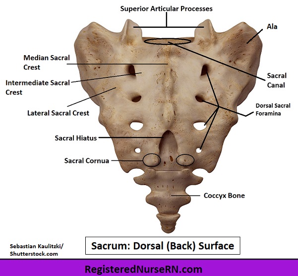
Dorsal Surface of the Sacrum
- Superior articular processes – At the superior (top) portion of the sacrum, there are two processes that look like horns. These are called the superior articular processes. These articulate (form a joint) with the fifth lumbar vertebra (L5), creating two zygapophyseal joints, which are synovial plane joints.
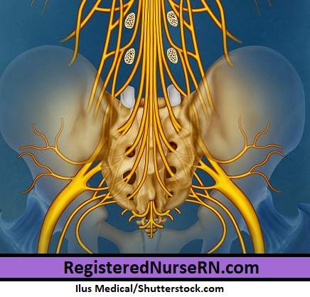
- Sacral canal – Between the superior articular processes is the sacral canal, which is a hollow opening that serves as a continuation of the vertebral canal above, and it houses the sacral nerves.
- Dorsal and ventral foramina – The sacral nerves will pass through the sacral canal and exit through four pairs of foramina (holes) on both the front (ventral) and back (dorsal) surfaces of the sacrum. The ventral foramina are on the front or anterior surface, and the dorsal foramina are on the back surface (posterior).
- Sacral hiatus – Because the inferior spinous process and laminae fail to develop on the lower portion of the sacral bones, there is an opening in the sacral canal that resembles a triangle, called the sacral hiatus. The word hiatus just means “gap,” so this is the gap on the inferior end of the sacral canal.
- Sacral cornua – The sides of the sacral hiatus terminate into the two sacral cornua. The word cornua comes from the Latin, and it means horn. So these two sacral cornua are going to articulate (form a joint) with the coccygeal cornua of the coccyx bone.
- Median, lateral, and intermediate sacral crests – There are three major crests on the dorsal (back) surface of the sacrum.
- As you look at the middle of the surface of the sacral canal, you’ll notice a bumpy ridge called the median sacral crest. These bumps are the remnants of the spinous process of each sacral vertebra, and they allow for the attachment of the supraspinous ligament. Remember: median is a directional term that means midline, so this is the crest in the midline region of the sacrum.
- The intermediate sacral crests run medial to the dorsal sacral foramina, and they allow for the attachment of the posterior sacroiliac ligaments. Remember that intermediate means between two structures, and this is between the median and lateral crests.
- And on the lateral (side) portion of the sacrum, you have the lateral sacral crest, which represent the remnants of the transverse process of each vertebra. This crest allows for the attachment of the sacrotuberous and sacroiliac ligaments. Remember, lateral means toward the side (or away from the midline) of the body.
Ventral Surface of the Sacrum
Now let’s look at the ventral or front (anterior) surface of the sacrum.

- Base – At the superior (top) region of the sacrum, you’ll notice an oval-shaped area, which is called the base. This allows for articulation with that fifth lumbar vertebra via its intervertebral fibrocartilage, creating the lumbosacral joint.
- Sacral Promontory – The first sacral vertebra’s body projects forward on its anterior side, creating the sacral promontory, a posterior portion of the pelvic inlet.
- Ala – The first sacrum’s body spreads out into a wing-like structure on each side called the ala (ala = wing; alae = plural). The alae allow for the attachment of muscles and ligaments, and at the side, they articulate with the ilium’s auricular surface, creating the sacroiliac joints.
- Lateral part – Inferior to the alae, there is a section of bone that is lateral to the foramina, which anatomists call the lateral part of the sacrum.
- Apex – Finally, we have the apex, which is the inferior end of the sacrum. Again, apex means “point,” so this is the end point, if you will, of this triangular bone. The apex articulates with the base of the coccyx bone, forming the sacrococcygeal symphysis, an amphiarthrodial joint that allows only slight movement.
Free Quiz and More Anatomy Videos
Take a free sacrum anatomy quiz to test your knowledge, or review our sacrum anatomy video. In addition, you might want to watch our anatomy and physiology lectures on YouTube, or check our anatomy and physiology notes.