In this anatomy lesson, I’m going to cover the patella bone, also known as the kneecap. The patella bone is part of the appendicular skeleton, and it gets its name from a Latin word that means “shallow pan or dish.”
Anatomists classify it as a sesamoid bone, which is often considered a subcategory of short bones, and it is the largest sesamoid bone found in the human skeleton.
Patella Function
Because the patella bone is located within the quadriceps tendon, it adds additional strength and leverage for those muscles, while also adding a layer of protection for the knee’s joint.
Patella Significance for Nurses and Healthcare Professionals
Nurses and other healthcare professionals will often use the patella bone as a landmark when checking the patellar reflex, as Nurse Sarah indicated in our video on the patellar reflex.
Patella Anatomy
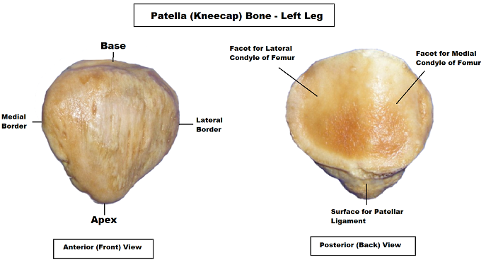
As you examine the anatomy of the patella, you’ll notice that it has a triangular shape that resembles a rounded arrowhead. In this picture we are looking at the anterior (or front) and posterior (back) view of the left patella bone.
Patella Anterior (Front) View
- Apex – The inferior (or bottom) portion of the patella comes to a point, which is called the apex (apex means point), and it always points toward your foot – so that’s how you’ll know the proper orientation of the bone. This apex allows for the attachment of the patellar ligament, which connects to the tibial tuberosity on the anterior (or front) surface of the tibia bone.
- Base – Toward the superior portion (or top border) of the patella, we have the base. This is going to allow for the attachment of the quadriceps femoris muscle group.
- Lateral and medial border – the left and right sides of the patella are called the lateral and medial borders. The lateral border allows for the attachment of the vastus lateralis, and the medial border allows for the attachment of the vastus medialis muscle.
Patella Posterior (Back) Surface:
When we look at the posterior (or back) view of the patella bone, we see that it has two large, smooth facets for the upper portion, as well as a rough surface toward the inferior portion.
- Surface for Patellar Ligament – This rough, inferior portion, which is called the surface for the patellar ligament, can help you determine that you’re looking at the posterior view of the patella. As the name suggests, part of the patellar ligament attaches to this rough surface.
- Facet for Lateral Condyle of Femur – Above this rough surface, you’ll notice the two smoother facets, which allow for articulation with the femur bone. If you think back to my video on the femur, I talked about the patellar surface of the femur, which was a depression that accommodates the patella bone.
- The lateral facet articulates with the lateral condyle of the femur, and it is the larger of the two facets. That will tell you whether you have a left or right patella bone. Just find the bigger facet, and you’ll know that the bigger facet represents the lateral side, which points away from the body’s midline. In this picture, we can tell that it is the patella of the left leg. If the larger facet is on the left side, it’s a left patella bone. If it’s on the right side (when looking at the posterior view), it’s the right patella bone.
- Facet for Medial Condyle of Femur – The medial facet is smaller and has a sharper angle, and it is going to articulate with the medial condyle of the femur bone. It’s always going to be positioned toward the midline of the body.
Free Quiz and More Anatomy Videos
Take a free patella anatomy quiz to test your knowledge. In addition, you might want to watch our anatomy and physiology lectures on YouTube, or check our anatomy and physiology notes.