The nail unit consists of the nail (fingernail or toenail), which is made of hardened plates of keratin, and its surrounding tissues that make up the distal, dorsal surface of the fingers and toes (digits). Nails are one of the appendages that, along with the skin, make up the integumentary system.
The other appendages of the integumentary system include the hair, sweat glands, and sebaceous (oil) glands.
Function of Nails
Our nails have several important functions. To help you remember them, use the mnemonic “STOP:”
- Sensory assistance – our nails provide input for the nerves in the underlying skin tissue.
- Tool functionality –nails allow you to do things such as dig around, scratch the skin, grasp objects, scrape objects, etc.
- Ornamental – nails act as a beauty accessory in many cultures, especially with the ladies who like to get their nails manicured.
- Protection – nails protect and support the distal ends of our digits as we use them for various tasks.
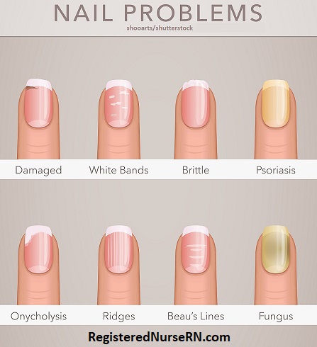
Nails Have Clinical Relevance
Nails also have an important clinical relevance, and healthcare professionals use them to assess the health of patients. For example, nurses may perform something called a capillary refill test by pressing on the nail plate, which temporarily blanches the underlying tissue. The nurse then releases the nail plate and observes the time it takes for blood flow to return to the nail bed, which can help assess for things such as dehydration or proper blow flow in the extremities.
In addition, unusual nail characteristics such as white bands, deep horizontal ridges, or discoloration may also point to underlying health problems.
Nail Anatomy
Let’s take a look at the anatomy of the nail unit, starting with the nail itself.
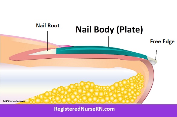
The Free Edge of a Nail
The free edge is the distal-most part of the nail that you have to trim with fingernail clippers. It often has a white appearance as it grows out, because this part of the nail is no longer attached to the underlying nail bed, which is what gives the nail body its pinkish color.

The Nail Body (Plate)
The nail body (or nail plate) makes up the hard, visible portion of the fingernail that attaches to the underlying epidermis. It is made of rows of dead keratinocytes that are translucent, allowing the color of the underlying tissue, which is rich in vessels, to show through.
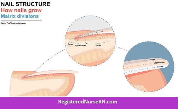
The plate itself can be divided into three main layers: a dorsal layer (top), an intermediate layer (middle), and a ventral layer (bottom).
The Nail’s Root
The nail’s root is the proximal-most portion of the nail that is hidden within the skin.
Underlying Nail Tissue
Now let’s look at the underlying tissues responsible for the creation and anchoring of the nail plate.
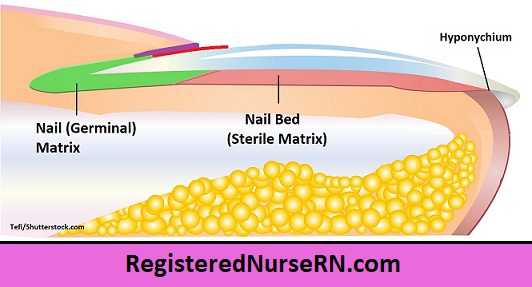
The Germinal Matrix (Nail Matrix)
The germinal matrix (or simply “nail matrix“) is under the skin and originates behind (proximal to) the nail’s root. The germinal matrix causes most of the nail’s growth by creating new cells that migrate forward. These cells eventually flatten and lose their nuclei in the process. This germinal matrix can extend all the way to the proximal part of the nail’s body and and is sometimes visible as the lunula.
The Lunula of the Nail
The lunula’s name means “little moon” (think lunula = lunar), and this is actually the distal end of the germinal matrix, which is not translucent like the nail’s body, so it has a different color. The lunula tends to be most visible on the thumb (pollex) or big toe (hallux). However, on other digits, the lunula may or may not be visible.
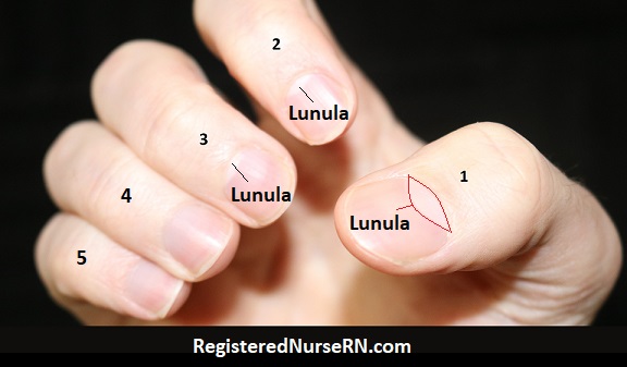
On my hand, the lunula is only visible on digits 1-3, but some people have them visible on all five digits of each hand or foot (or even none at all), which can be normal. However, the lunulae (plural) may also disappear from your digits entirely if you have an underlying disease or experience organ failure, such as kidney failure. [1]
Nail Bed (Sterile Matrix)
The nail bed (or sterile matrix) is the area found between the lunula of the nail matrix and the hyponychium, and this tissue allows for the attachment of the nail body (plate). The dermis and epidermis of the nail bed often form into longitudinal ridges, and sometimes you can see small vertical ridges on the nail plate itself. These ridges can be normal. However, deeper ridges or horizontal ridges in the nails could indicate an underlying health issue.
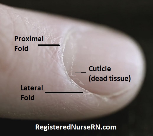
Nail Folds
There are also skin folds that surround the nail plate.
- The proximal fold is the fold of skin near the proximal end (origination) of the nail plate.
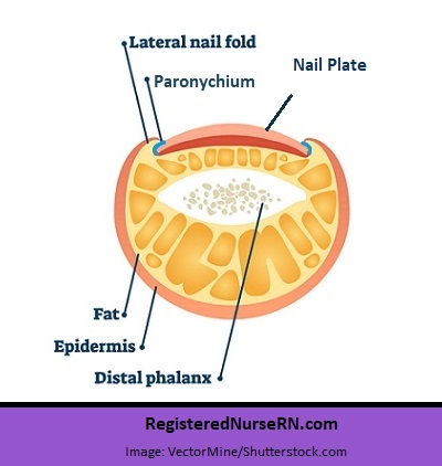
- The lateral folds lay on each side of the nail plate.
Note: In some texts, the proximal fold may be considered the same as the eponychium, and the lateral fold may be referred to as the paronychium. See below for details of the eponychium and paronychium.
Nail Unit Tissue (Hyponychiu, Eponychium, Paronychium)
Finally, we have special tissues around the nail folds that help seal and protect the nail plate. These words all end in “nychium,” which means “little claw,” referring to the nail itself. The prefix for each word tells you where these tissues are located in relation to the nail.
Hyponychium
The hyponychium is the epithelial tissue just under the free edge of the nail and distal to the nail bed. Remember, the prefix “hypo” means “under”, so it’s under the “little claw.” The hyponychium is rich in white blood cells and forms a seal between the nail plate and nail bed to defend against germs. The onychodermal band is present where the hyponychium joins the nail bed.
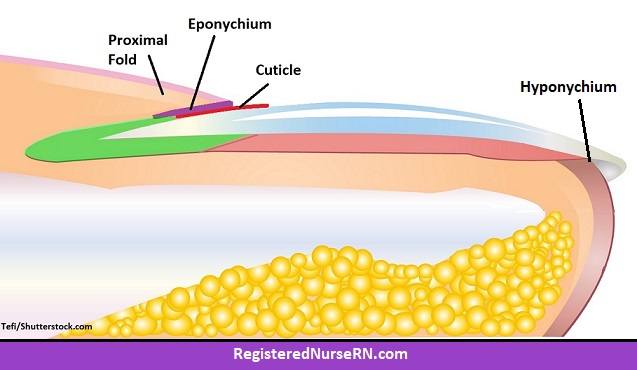
Eponychium
The eponychium is the thin layer of tissue on the ventral surface of the proximal fold that covers, seals, and protects the nail’s matrix and root. The prefix “epo” or “epi” means “over” or “upon”, and again “nychium” refers to nail/claw, so this is over or upon the nail at its root. The eponychium produces the cuticle.
Cuticle of the Nail
The cuticle is the thin, dead, transparent tissue produced by the eponychium, and it extends onto the base of the nail plate. Note: You may also see the eponychium referred to as the cuticle of the nail (or even the proximal fold in some texts or illustrations), but these are technically different. (For testing purposes, follow whatever your teacher or textbook suggests.)
Paronychium
The paronychium is the tissue along the border of the nail plate at the lateral folds. The prefix “paro” or “para” means alongside or beside, and “nychium” refers to the nail. If you get an infection here it’s called paronychia. Note: Some texts may treat the lateral fold and the paronychium as one and the same.
Perionychium
Finally, the perionychium is a word that describes all of the tissues that surround the nail’s body (peri means all around), including the paronychium, eponychium, hyponychium, lateral and proximal folds, and the nail bed and matrix.
Free Quiz and More Anatomy Videos
Take a free quiz on nail anatomy to test your knowledge, or review our nail anatomy video. In addition, you might want to watch our anatomy and physiology lectures on YouTube, or check our anatomy and physiology notes.
References:
1. Daadaa N, Souissi A, Ben Kaab B, Zouaghi MK, Mokni M. Absent lunula: An overlooked finding in chronic kidney disease. Clin Case Rep. 2020 Nov 20;9(1):576-577. doi: 10.1002/ccr3.3471. PMID: 33489220; PMCID: PMC7813076.