- Basics about Stethoscope Usage
- Basics about Heart Sounds
- Auscultation Sites
- Patient Positioning
- Tips for Distinguishing S1 from S2
- Anatomy of the heart and blood flow (video)
- Basics about S3, S4, and heart murmurs
Lecture on Heart Sounds
Purpose of Heart Auscultation
Goal: to assess the closure of the heart valves (Aortic, Pulmonic, Tricuspid, Mitral “Bicuspid”)Basics about Stethoscope Usage
Before you listen to heart sounds you must know how to use your stethoscope’s chest piece properly.- Diaphragm: use for listening to HIGH PITCHED sounds like S1, S2, and aortic/pulmonic murmurs
- Bell: use for listening to LOW PITCHED sounds like S3, S4, and mitral stenosis murmurs
ECG/EKG Study Guide and Workbook for Nursing Students

You can get an eBook version here: “Nurse Sarah ECG Book” or a physical copy here: “ECG/EKG Interpretation Study Guide by Nurse Sarah“.
Basics about Heart Sounds
Heart sounds are caused by the closure of heart valves. The first sound you hear is S1 and is caused by the closure of the atrioventricular valves (AV) TRICUSPID AND MITRAL VALVES. This sounds like “LUB”. The second sound you hear is S2 and is caused by the closure of the semilunar valves (SL) AORTIC AND PULMONIC VALVES. This sounds like “DUB”. Normally, the AV valves close at the same time and the same is true for the SL valves. However, in some people these valves may close asynchronously and this would cause a split in sound.- S1 split: Tricuspid and mitral valves closing asynchronously
- S2 split: Aortic and pulmonic valves closing asynchronously
Heart Auscultation Sites
Remember the mnemonic “All Patients Take Medicine”. This represents the order of how you will auscultate the heart. Memorize the information below because it is crucial in understanding heart sounds and which valve represents each sound.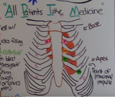 Aortic: found right of the sternal border in the 2nd intercostal space REPRESENTS S2 “dub”
Pulmonic: found left of the sternal border in the 2nd intercostal space REPRESENTS S2 “dub”
Erb’s Point: found left of the sternal border in the 3rd intercostal space
Tricuspid: found left of the sternal border in the 4th intercostal space REPRESENTS S1 “lub”
Mitral: found left of the sternal border at the midclavicular in the 5th intercostal space REPRESENTS S1 “lub” (also the site of point of maximal impulse)
The Base of the heart includes the aortic and pulmonic areas, and S2 will be loudest at the base. Aortic and pulmonic murmurs are heard best at the base with the patient leaning forward and sitting up with the diaphragm of the stethoscope.
The Apex of the heart includes the tricuspid and mitral areas, and S1 will be loudest at the apex. S3 and S4 along with mitral stenosis murmurs will be heard best at this position with the patient lying on their left side with the bell of the stethoscope.
Aortic: found right of the sternal border in the 2nd intercostal space REPRESENTS S2 “dub”
Pulmonic: found left of the sternal border in the 2nd intercostal space REPRESENTS S2 “dub”
Erb’s Point: found left of the sternal border in the 3rd intercostal space
Tricuspid: found left of the sternal border in the 4th intercostal space REPRESENTS S1 “lub”
Mitral: found left of the sternal border at the midclavicular in the 5th intercostal space REPRESENTS S1 “lub” (also the site of point of maximal impulse)
The Base of the heart includes the aortic and pulmonic areas, and S2 will be loudest at the base. Aortic and pulmonic murmurs are heard best at the base with the patient leaning forward and sitting up with the diaphragm of the stethoscope.
The Apex of the heart includes the tricuspid and mitral areas, and S1 will be loudest at the apex. S3 and S4 along with mitral stenosis murmurs will be heard best at this position with the patient lying on their left side with the bell of the stethoscope.
Patient Positioning for Heart Auscultation
- Supine or sitting-up: Use the diaphragm and listen at all 5 auscultation sites (noting S1 and S2 and if there are any splits presents). In addition, distinguish S1 from S2. Then repeat with the bell of the stethoscope…noting any other extra sounds.
- Left side: turn the patient onto their left side and auscultate with the bell of the stethoscope at the APEX area and listen for S3, S4, or mitral stenosis murmurs.
- Sit up, lean forward, and have patient exhale: Listen with the diaphragm at the aortic and pulmonic sites for murmurs.
Tips for Distinguishing S1 from S2
This is complicated when first starting out with heart auscultation, but it’s vitally important so you can assess extra heart sounds.- S1 is louder at the apex.
- Feel on the carotid artery while listening at the apex (S1 correlates with the carotid pulsation).
- EKG monitor: if the patient is on a bedside monitor the r-wave of the QRS complex will correlate with the sound of S1.
Basics about S3, S4, and Heart Murmurs
S3 and S4 are heard best at the apex of the heart with the bell of the stethoscope while the patient is on their left side. Note: it is normal for a patient NOT to have a S3, S3, or heart murmur- S3: heard after S2 and sounds like “LUB-DUB-TA”
- Caused by vibrations of ventricle filling from a resistant ventricle due to fluid volume overload or heart failure.
- S4: heard before S1 and sounds like “TA-LUB-DUB”
- Caused by ventricle resistance from an atrial “kick” during presystole (hypertrophic left ventricle)
- Heart murmurs: blowing/swooshing noise from blood turbulence in the chambers of the heart (wall defect) or valve problem (stenosis or regurgitation)
- Grade 1: hard to hear
- Grade 2: faint but heard
- Grade 3: easily to hear
- Grade 4: Loud with a chest thrill
- Grade 5: Very loud…can hear when corner of the chest piece is lifted off the chest
- Grade 6: Loudest…can hear when whole chest piece lifted off the chest
- **Note Grades 4-6 a thrill is present
- -Base on Levine Scale