Arterial vs. venous ulcers nursing review that covers the differences between these two types of lesions that can occur when a patient has peripheral vascular disease.
As a nursing student or nurse, you must be familiar with these types of ulcers. What should you know for exams? It’s important to know the location variations between arterial and venous ulcers along with their defining characteristics.
Arterial vs. Venous Ulcers Nursing Review
Arterial Ulcers vs. Venous Ulcers
Arterial:
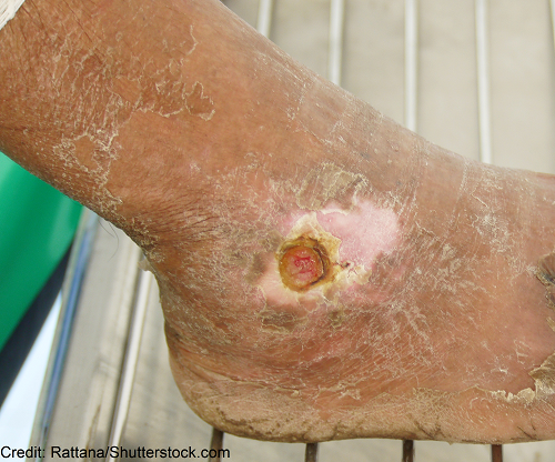
Location: end of toes, top of feet (dorsum), lateral ankle region (lateral malleolus)
Ulcer’s Appearance?
- Very little drainage
- Little tissue granulation (pale/very light pink) OR necrotic/black
- Deep “punched out” w/ noticeable margins/edges that gives it a round appearance
Click Here to see -> Arterial Ulcer…note the punched out appearance, how it is located on the lateral malleolus, it has little drainage, and the wound base is very pale (in addition, the surrounding skin is very scaly/dry).
Venous:
Location: medial parts of lower legs & medial (malleolus) ankle region
Ulcer’s Appearance?
- Swollen w/ drainage
- Granulation present (deep pink to red)
- Edges are irregular and depth is shallow
Click Here to see -> Venous Stasis Ulcer…note it is located on the medial part of the lower leg and medial malleolus, wound base is a deep red color, edges are irregular, and the skin surrounding the wound is tight and has edema and brown pigmented.
You may be interested the peripheral vascular disease review.
References:
Peripheral Artery Disease (Also Known as P.A.D.) Retrieved 4 October 2019, from https://www.nhlbi.nih.gov/health-topics/peripheral-artery-disease
Treatment Strategies for Patients with Lower Extremity Chronic Venous Disease (LECVD). Retrieved 4 October 2019, from https://www.cms.gov/medicare-coverage-database/details/technology-assessments-details.aspx?TAId=104&bc=AAAQAAAAAAAAAA%3d%3d&