 Now let’s break each part of this strip down and talk about what each area represents. You always read the PQRST wave starting from left to right. Please take note of the study tips because these are common questions are exams.
Basic PQRST:
P-wave: The first little “hump” or “bump” you see is known as the P-wave. Remember from the electrical conduction lecture, that the SA node is responsible for this.Study tip: The P-wave represents ATRIAL DEPOLARIZATION (depolarization is a big, fancy word for CONTRACTION).
Study tip: The QRS complex represent VENTRICLE DEPOLARIZATION (contractions of the ventricles)
T-wave: After this spike, you will see a “bump” shortly after the complex. This “bump” is called the t-wave and is caused by the ventricles relaxing. The ventricles are so large that when they contract (depolarize) the form a large electrical impulse that presents the QRS complex. Therefore, (because they are so large) when they relax (repolarize) they form a small electrical impulse that presents as the t-wave.Study tip: What area of PQRST EKG reading represents ventricle repolarization? T-wave
U-wave: This is not very common, but I wanted to show it to you and mention it. The u-wave sometimes is seen after the t-wave. This is thought to be caused by the relaxation of the Purkinje fibers.ECG/EKG Study Guide and Workbook for Nursing Students
You can get an eBook version here: “Nurse Sarah ECG Book” or a physical copy here: “ECG/EKG Interpretation Study Guide by Nurse Sarah“.
PR Interval & ST Segment:
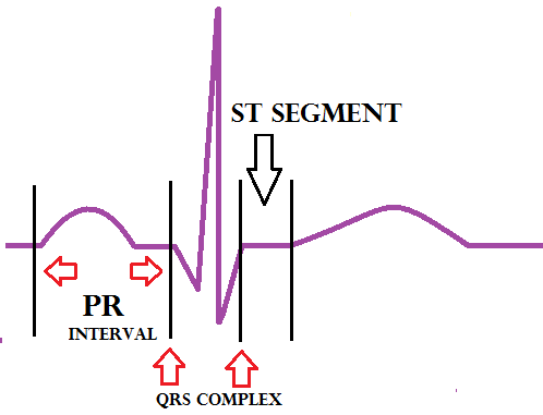 PR -interval: As noted on the diagram above, the PR-interval starts at atrial contraction (remember atrial contraction is represented by the P-wave) and ends at the beginning of ventricle depolarization. So in other words, it starts at the P-wave and ends at the beginning of the QRS complex.
ST segment: This segment starts at the J-point. The J-point is where you start to see an upward stroke after the S wave. The segment ends at the beginning of the T-wave. The ST-segment represents when the ventricles are relaxing, also called repolarizing.
Now test your knowledge on how well you grasp the material by taking this quiz. If you would like more explanations or are a visual learner, I suggest you check out this teaching tutorial.
PR -interval: As noted on the diagram above, the PR-interval starts at atrial contraction (remember atrial contraction is represented by the P-wave) and ends at the beginning of ventricle depolarization. So in other words, it starts at the P-wave and ends at the beginning of the QRS complex.
ST segment: This segment starts at the J-point. The J-point is where you start to see an upward stroke after the S wave. The segment ends at the beginning of the T-wave. The ST-segment represents when the ventricles are relaxing, also called repolarizing.
Now test your knowledge on how well you grasp the material by taking this quiz. If you would like more explanations or are a visual learner, I suggest you check out this teaching tutorial.