This NCLEX review will discuss kidney and nephron anatomy.
As a nursing student, you must be familiar with the basic structure of the kidney and its nephron.
It will help you understand the diseases that affect the renal system.
Don’t forget to take the kidney and nephron anatomy quiz.
You will learn the following from this NCLEX review:
- Kidney anatomy
- Nephron anatomy
- How urine is produced
- How urine flows out of the kidneys
Lecture on Kidney and Nephron Anatomy
Kidneys and Nephron Anatomy NCLEX Review
What is the role of the kidneys? In short, to filter the blood. The kidneys produce urine with the assistance of the nephron. The urine contains waste and excessive water/minerals we don’t need. The kidneys want to maintain a fine balance of water and electrolytes in the body and remove excessive waste.
 Key Concepts about the Kidneys:
- You have two kidneys (right and left).
- The right kidney sits lower than the left kidney. WHY? To help accommodate the large size of the liver, which sits right above the right kidney.
- The kidneys receive fresh blood from the heart via the renal artery, and drains filtered blood back to the heart via the renal vein.
- The nephron is the functional part of the kidney….meaning it is where urine is produced.
- Each kidney contains MILLIONS of nephrons which are found in the renal cortex and renal medulla of the kidney.
- The nephron consists of the following parts:
- Renal Corpuscle (function is to FILTER the blood and create filtrate)
- Glomerulus
- Bowman ’s capsule
- Renal Tubule (function is to REABSORB and SECRETE substances IN or OUT of the filtrate with the assistance of the peritubular capillaries)
- Proximal Convoluted Tubule
- Loop of Henle
- Distal Convoluted Tubule
- Collecting Tubule
- Renal Corpuscle (function is to FILTER the blood and create filtrate)
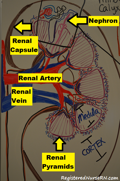
Basic Parts of the Kidney:
Renal Capsule: outer layer of the kidney that gives the kidney its shape and protects the kidney from infections that may occur from surrounding organs.
 If you slice the kidney in half you will see:
Renal cortex: outside layer that contains the renal corpuscles (glomerulus and bowman’s capsule which primary functions are to FILTER the urine) and renal tubules EXCEPT the loop of HENLE ( it is found in the renal medulla).
Renal medulla: inside layer (found within the renal pyramids) which is very hypertonic “salty”. These conditions help maintain water and salt balance in our body with the help of the nephron, specifically the Loop of Henle.
Blood supply:
Renal artery: takes fresh oxygenated blood that came from the heart to the kidney to be filtered. It further branches off around the renal columns into the renal cortex into arterioles (afferent/efferent) then peritubular capillaries.
Renal vein: take filtered blood back to the heart to be re-oxygenated and pumped to the body. It comes from the efferent arterioles.
Renal pyramids: found within the renal medulla which contains the loop of henle and parts of the collecting tubule.
Renal papilla: pointed projection of the renal pyramid that project into the minor calyx and major calyx
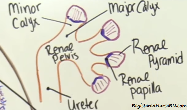 Minor and Major calyx along with the renal pelvis, ureters, bladder, and urethra play a role in draining out the urine that has left the nephron.
Nephrons: the functional part of the kidneys that filters the blood (renal corpuscle), reabsorbs minerals/water and secretes waste (renal tubule), and produces the substance called urine which will drain down into the ureters, be stored in the bladder, and voided out via the urethra.
—In the renal cortex and medulla: Parts of the nephron sit in the renal cortex, specifically the glomerulus and bowman’s capsule, proximal convoluted tubule. It then dips down in the renal medulla specifically the loop of henle. Then the distal convoluted tubule goes back up into the renal cortex along with parts of the collecting tubule which then goes back down into the renal medulla and connects directly to the renal papilla and urine excretion begins.
Individual role of each part of the nephron:
 How does the nephron receive its blood supply?
From the AFFERENT ARTERIOLE…the afferent arteriole sends blood to the first part of the nephron called the Glomerulus.
Glomerulus: It is a collection of circular capillaries that have extremely high pressure which help perform ULTRAFILTRATION. During this process the blood will be filtered and FILTRATE will be created (which is a liquid consisting of the collection of fluid and particles that came from the blood). These substances will “drip” down into a capsule that surrounds the glomerulus called BOWMAN’S CAPSULE (collects the filtrate).
What drips down?
- Water
- Ions: sodium, chloride, calcium, potassium, magnesium, phosphate, bicarbonate
- Amino acids
- Glucose
- Creatinine
- Urea
****not filtered blood cells or proteins
Then the newly filtered blood exits via the EFFERENT arterioles which will go on and form the peritubular capillaries that will surround the nephrons. The peritubular capillaries on the loop of henle are known as the vasa recta.
The peritubular capillaries will play a role in carrying the reabsorbed nutrients from the filtrate back into the body’s system to the renal vein and secreting substances (urea, ions) and drugs found in the blood into the tubules at certain points.
Side Note….why do we called it RE-absorption rather the just absorption? The substances filtered from the glomerulus where already ABSORBED at some point in the GI tract via specialized cells which took the nutrients into the blood stream (most of this happens in the small intestine). Then the substances traveled through the body via the heart and made their way to the kidneys via the renal artery to be FILTERED out. Therefore, our body will RE-ABSORB these nutrients based on what our body needs (because we already absorbed them once from the food we ate). Then the left overs will be excreted in the urine.
The created filtrate then flows through the proximal convoluted tubule (PCT) and this tubule reabsorbs MOST of the parts of the filtrate that we need to survive which just came from the Bowman’s capsule.
Then the filtrate enters into the Loop of Henle ( remember it is found down in the renal medulla). The loop of henle has a descending limb and ascending limb. Its goal is to concentrate the urine and it will accomplish this with the renal medulla. The renal medulla’s interstitial fluid is very hypertonic. This helps reabsorb water from the filtrate to maintain the body’s water and salt balance.
The descending limb is ONLY PERMABLE TO WATER…while the ascending limb is ONLY PERMABLE TO IONS.
The filtrate then enters in the distal convoluted tubule where more substances are reabsorbed and secreted. Then it travels to the collecting tubule where the filtrate is brushed up with the final touches of reabsorption. Then the filtrate leaves the collecting tubule as urine which again flows through the renal papilla, minor/major calyx, renal pelvis, ureters, bladder, and urethra.
References:
- Marieb, Elaine Nicpon, and Jon Mallatt. Human Anatomy. 3rd ed. San Francisco: Benjamin Cummings, 2001. Print.
- “Your Kidneys And How They Work | NIDDK”. National Institute of Diabetes and Digestive and Kidney Diseases. N.p., 2014. Web. 25 Apr. 2017.