In this anatomy lesson, I’m going to cover the anatomy of the tibia and fibula bones of the anatomical leg, which is the section between the knee and ankle. These two leg bones are part of the appendicular skeleton, and anatomists classify them as long bones.
Tibia and Fibula Memory Trick
If you get these two bones mixed up, here’s a quick memory trick: remember the phrase “never tell a little fib.” The fibula is the smaller of the two bones, so “little fib” will help you remember that it is the small one. Also, little starts with the letter “l,” which can help you remember that this bone is always on the lateral side.
Interosseous Membrane
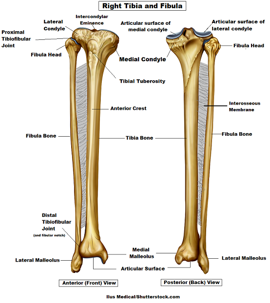
First, you’ll notice this web-like structure called the interosseous membrane (or middle tibiofibular ligament). The prefix “inter” means between, and osseous refers to the bones. So this is literally the membrane between the bones. (Note, the radius and ulna bones also have this membrane.)
This membrane keeps the tibia and fibula together and provides strength and stability for them. It also separates muscles on the anterior and posterior parts of the leg.
Tibia Bone Anatomy
Now let’s look at the tibia bone, which is the larger of the two leg bones, located medially. When you feel your shinbone, this is what you’re feeling. In fact, this bone gets its name from a Latin word that literally means “shinbone.”
The tibia’s larger size allows it to transfer weight from the femur to the foot.
- Medial and lateral condyles – The proximal end (or top) of the tibia widens out from the shaft and forms a medial and lateral condyle. A condyle is a rounded projection on a bone that is going to articulate (or form a joint) with something. Medial is a directional term that tells us it is toward the midline of the body, and lateral means away from the body’s midline.
- Articular surface of medial and lateral condyles – The top of these condyles have a concave surface, which you can see on the posterior view of the tibia. These are called the articular surface of the medial and lateral condyles. This is going to articulate with the medial and lateral condyles of the femur (thigh) bone, forming the tibiofemoral joint.
- Intercondylar eminence – Between the articular surface of the medial and lateral condyles is an area called the intercondylar eminence. This structure includes a left and right tubercle, which resembles tiny devil horns, and fossae, to which the menisci and the cruciate ligaments attach.
- Tibial tuberosity – Between the condyles and the shaft, you’ll notice a bump on the anterior side of the tibia called the tibial tuberosity. That’s all a tuberosity is: it’s a fancy word that means bump or small rounded area. This structure allows for the attachment of the patellar ligament.
- Anterior crest – Below the tibial tuberosity we have the triangular shaft of the tibia, which contains three borders and three surfaces. You’ll notice that the anterior side of the tibial shaft forms a pointed border called the anterior crest. This area forms the hard shin area of the leg and allows for the attachment of the deep fascia.
- Fibular notch – Finally, we have the fibular notch, which is a depression that allows for the attachment of the fibula bone, forming the distal tibiofibular joint.
- Articular surface of the tibia – The distal end of the tibia will transfer weight to the foot at its articulation with the talus bone, forming the ankle joint.
- Medial malleolus – On the medial side of the tibia’s distal end, there is a rounded bony area with a projection called the medial malleolus. Malleolus comes from a Latin word that means “little hammer,” and this area articulates with the talus bone of the foot. This is the bony area you feel on the inside of your ankle. It can serve as a landmark when locating the posterior tibial pulse point.
Fibula Bone Anatomy
Unlike the tibia bone, the fibula is not a weight-bearing bone. However, it allows for the attachment of various muscles. Below are some of the important structures on the fibula bone.
- Head – Starting at the proximal (top) end of the fibula, you’ll notice the larger head on the fibula. This is going to articulate (form a joint) with the lateral condyle of the tibia bone, forming the proximal tibiofibular joint. It also provides attachment for the biceps femoris and fibularis longus, as well as various ligaments.
- Shaft – The shaft of the fibula is thin and ridged, which allows for the attachment of various muscles of the leg.
- Lateral malleolus – Finally, we have the lateral malleolus, which forms the bony part of the outer (lateral) ankle. Like the medial malleolus, this articulates (forms a joint) with the talus bone of the foot (see foot bones) and allows for the attachment of ligaments. Although the fibula bone is smaller and thinner than the tibia, the lateral malleolus of the fibula is larger than the medial malleolus of the tibia.
Free Quiz and More Anatomy Videos
Take a free tibia and fibula anatomy quiz to test your knowledge. In addition, you might want to watch our anatomy and physiology lectures on YouTube, or check our anatomy and physiology notes.