Cardiogenic shock nursing NCLEX review for students!
In this review you will learn about cardiogenic shock.
After reviewing these notes, don’t forget to take the quiz that contains cardiogenic NCLEX questions and to watch the lecture.
Lecture on Cardiogenic Shock
Cardiogenic Shock NCLEX Review
What is cardiogenic shock? It’s where the heart can NOT pump enough blood to meet the perfusion needs of the body.
 Therefore, the heart does not pump enough blood throughout the body, which will decrease cardiac output and this leads to a decrease in tissue perfusion and oxygen supply to the organs/tissue’s cells.
Note: In cardiogenic shock, there is NOT an issue with a loss of blood volume like in some of the other types of shock. Blood volume is normal. However, because the heart has experienced a decrease in its ability to function properly, that blood volume starts to back up and leads to congestion in the lungs and the right side of the body (keep this in mind for when we review the signs and symptoms).
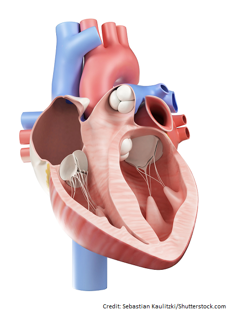
Let’s break down the heart’s role:
The heart is the PUMP of the body. It takes blood to the right side of the heart that has been used and depleted of oxygen by the cells of the body and pumps it to the lungs. The lungs oxygenate and remove carbon dioxide from the blood and sends it back to the heart via its left side.
The left side is very strong, especially the left ventricle, which is the main pumping chamber of the heart. The left side shoots the fresh oxygenated blood into the aorta. The aorta branches off into many arteries that go into this complex network of vessels to feed the organs oxygenated blood.
Therefore, the heart is the main center piece in the body that plays a role in tissue perfusion, and everything in the body depends on the heart for its survival. If the heart can’t pump blood efficiently, the organs don’t receive fresh oxygenated blood and the cells that make up that organ start to die.
Therefore in a nutshell, when the heart can’t pump, cardiac output falls, which decreases tissue perfusion and the cells that make up our organs don’t receive enough oxygen to work. They start to panic and eventually die.
When cardiac output falls (hence the blood pressure drops), the patient begins to enter the stages of shock (we talked about the stages of shock in the previous lecture: initial, compensatory, progressive, and refractory).
Cardiac Output and Cardiogenic Shock
First, let’s talk about cardiac output because it’s a very important term you should know when taking care of a patient with cardiogenic shock:
Cardiac output is the amount of blood the heart pumps per minute. The heart’s cardiac output should be anywhere from 4-8 liters of blood per minute.
Cardiac output is determined by the person’s heart rate times the stroke volume.
Stroke volume is the amount of blood pumped from the left ventricle each beat (50-100 ml). Stroke volume is determined by the preload, afterload, and contractility of the heart.
Preload is the amount the ventricle stretches at the end of diastole (hence it’s the amount the ventricles stretch once their filled with blood….so it’s the end-diastolic volume). Remember diastole is the relaxation/filling phase of the heart.
Afterload is the pressure the ventricle must pump against to squeeze blood out. Therefore, it’s the force the heart has to pump against to get blood out of the ventricle.
Contractility is how well the muscle cells are contracting. Therefore, it’s how well the heart is contracting to pump blood.
If the stroke volume falls (meaning the heart muscle is NOT pumping blood very well), the cardiac output decreases and this leads to a fall in tissue perfusion.
So, in other words, cardiac output tells us how well the heart is pumping and perfusing the cells that make up our organs.
When we talk about treatment for cardiogenic shock it will include medications that will affect preload, afterload, and contractility via drugs like vasopressors, inotropes, vasodilators, and diuretics.
Examples:
- if the afterload is decreased (which is the resistance the heart must pump against to get blood out of the heart….if you decrease this, it makes it easier for the heart to pump blood forward) the stroke volume will increase hence delivering a higher cardiac output….vasodilators can achieve this.
- if contractility is increased (which is how strong the muscle cell contracts) it will increase stroke volume and cardiac output….positive inotropic medications can achieve this.
- if preload is increased (which is the amount the ventricle stretches at the end of diastole) the stroke volume will increase hence delivering a higher cardiac output… vasopressors can achieve this by causing vasoconstriction, which will increase venous return to the heart.
Another term you may see, especially when talking about cardiogenic shock, is Cardiac Index.
This is a more specific cardiac output measurement based on the patient’s body size. It’s calculated by taking the cardiac output and dividing it by the patient’s body surface area.
Patients with cardiogenic shock will have a CI (cardiac index) less than 2.2 L/min/m2.
Normal CI is: 2.5-4 L/min/m2
Causes of Cardiogenic Shock
What can cause the heart’s cardiac output to fall (hence cardiogenic shock)?
Issues that cause the heart to have problems:
- contracting (systolic dysfunction)…the heart can’t successfully pump blood out of the heart and to the body
- filling (diastole dysfunction)….the heart’s chambers don’t fill properly so blood really can’t be pumped to perfuse the body
Examples:
- ****Main cause: Acute myocardial infarction (contracting issue): this is known as a heart attack. It occurs due to a blockage in the coronary arteries. The coronary arteries supply the cells of the heart muscle with oxygen. When blood flow is dramatically decreased or blocked completely to the cells that make up the heart muscle, they start to die. When majority of the blood flow to the left coronary is diminished, the muscle cells that make up the left ventricle die. Remember the left ventricle is the main pumping chamber of the heart. It pumps the blood to the aorta which then goes to the body. If a patient has an acute myocardial infarction that affects the left ventricle, the patient is at high risk for developing cardiogenic shock.
- Why? When the heart cells die, the heart doesn’t contract (pump) like it should, especially the left ventricle. Preload, afterload, contractility are all going to be affected along with the heart rate. Cardiac output falls (can’t pump much blood per minute) and tissue perfusion is decreased to the body’s cells.
- Pericardial tamponade (filling issue): too much fluid is around the heart, so it can’t fill properly.
Other causes: dysrhythmias, myocarditis, endocarditis, pulmonary edema, structure issues with valves and/or septal walls etc.
Quick Patho on what happens during cardiogenic shock
Cause (ex: acute myocardial infarction) -> major cell damage to the heart muscle -> can’t pump efficiently…..ability to pump blood out of the heart and to the body has decreased -> decreases stroke volume -> decreased cardiac output -> decreases perfusion to the body’s cells that make up our organs and tissues -> cell injury (signs and symptoms of body systems struggling due to low oxygen)
Signs and Symptoms of Cardiogenic Shock
When trying to recall these signs and symptoms, think about what happens in the body due to decreased cardiac output and when blood starts to back flow into the lungs and the right side of the heart.
Let’s break down the signs and symptoms by body systems:
Heart: it’s weak and not pumping blood forward or filling efficiently…..this leads to:
- Back flow of blood from the left side to the lungs (pulmonary congestion, crackles, dyspnea, increased respiratory rate, low oxygen, increased heart rate)
- As it progresses, blood will back flow from the lungs to the right side of the heart (neck veins become distended due to an increase in venous pressure and there will be a high CVP…central venous pressure)
- Chest pain (decrease perfusion to heart muscle via the coronary arteries)
- Hypotension: Systolic blood pressure less than 90 mmHg or 40 mmHg from baseline for at least 30 minutes
- Weak peripheral pulses
- If on hemodynamic monitoring may have:
- Decreased cardiac index <2.2 L/min/m2
- Increased pulmonary capillary wedge pressure (PCWP) >18 mmHg
- Increased central venous pressure
Brain: cells are not being perfused due to low cardiac output and the build-up of waste in the body (liver and kidneys are failing to filter out toxic substances)…..leads to confusion, agitation, restlessness
Kidneys: decreased blood flow to the kidneys activates the renin-angiotensin system. This leads to the release of angiotensin II, which leads to the release of aldosterone and ADH (antidiuretic hormone). These substances will cause the kidneys to keep sodium and water (aldosterone) and cause the body to retain water (ADH)…..leads to oliguria <30 mL/hr urinary output
- WHY? These substances are naturally released by the body to help increase blood volume so cardiac output can be increased.
- Other signs and symptoms will be associated with renal failure like an increased BUN and creatinine.
Skin: decreased perfusion to the skin….leads to decreased capillary refill, cool, pale, and clammy skin
Nursing Interventions for Cardiogenic Shock
To understand the nursing interventions, we need to know the goals of medical treatment for a patient with cardiogenic shock. Goals include:
- reperfusion to the heart muscle if there is a decreased blood flow to the heart muscle via the coronary arteries. The can be performed via a heart catheterization, and if a blockage is found, a stent can be placed.
- increase cardiac output via cardiac medications (most meds are titrated and the patient is on hemodynamic monitoring to assess certain pressures in the heart)
- decrease fluid overload, if present (via diuretics)
- maintain oxygen status due to pulmonary edema (will need mechanical ventilation)
Nurse’s Role: Hemodynamic monitoring (more about this below), maintain mechanical ventilation (will need to help with respiratory failure), central line placement for medication, monitoring, assessing for signs of adequate tissue perfusion: mental status, blood pressure, heart rate, rhythm, urine output, skin color and capillary refill, lung sounds etc.
Monitoring labs: high cardiac markers ex: elevated troponin (this substance is released if there is injury to heart cells), increased BNP (this substance is released by the cells that make up the ventricles when they stretch due to high blood volume), pulmonary edema on chest x-ray, echocardiogram (show a low ejection fraction), acid-base level that demonstrate acidosis serum lactate >4 mmol/L (cells will switch from aerobic to anaerobic metabolism which will produce lactic acid), drop in blood ph <7.35
Hemodynamic monitoring:
Pulmonary artery wedge pressure/pulmonary capillary wedge pressure (increased in cardiogenic shock): a pulmonary artery catheter (Swan-Ganz catheter) is inserted through the right side of the heart via a vessel and the catheter is wedged in the pulmonary arterial branch and a balloon is temporarily inflated to measure left atrial filling pressure.
- Normal 4-12 mmHg
- >18 mmHg in cardiogenic shock….blood is backing up in the heart and lungs (hence not being pumped out and creating a high pressure in the left atrium)
Central venous pressure (CVP): catheter used to measure the pressure in the right atrium and superior vena cava.
- INCREASED in cardiogenic shock (Normal CVP is 2 to 6 mmHg or 8-12 mmHg depending on source).
- WHY? The back flow of blood to the right side of the heart (right atrium into venous circulation) leads to venous congestion, which increases the central venous pressure.
Medications: a combination of medications can be given and medications vary depending on the cause of the cardiogenic shock and the patient’s response.
Diuretics: cardiogenic shock is one of the two types of shock that doesn’t have a decrease in blood volume (the other is neurogenic shock).
- Patients with cardiogenic shock have a backup of fluid in the lungs from an injured heart that is failing to pump blood forward.
- Diuretics (ex: furosemide….watch potassium level “hypokalemia“….normal level is 3.5-5 mEq/L) will help remove extra fluid volume via the kidneys. This will decrease the preload (amount ventricles stretch at the end of diastole), which will help the relieve the workload of the heart. However, watch the fluid status of the patient, renal function, and for worsening hypotension.
Vasopressors: causes constriction of vessels and increases preload (venous return) and cardiac output
- Norepinephrine (vasoconstrictor) sometimes used over dopamine because it doesn’t cause tachydysrhythmias: it increases tissue perfusion by increasing blood pressure
Vasopressors that create a positive inotropic effect: increases the strength of the heart’s contraction (increases contractility) and stroke volume
- Dobutamine (increases contractility and cardiac output BUT can cause vasodilation due to the way it acts on receptors and may make hypotension WORSE. If this occurs that patient may be started on norepinephrine or dopamine.
- Dopamine (increase contractility, causes vasoconstriction: increases blood pressure and MAP (want >60 mmHg), but watch out for tachydysrhythmias)
Vasodilators: decreases preload and afterload: increases stroke volume because it decreases the afterload, so it’s easier for the heart to pump blood forward so cardiac output increases. The workload on the heart decreases by dilating the coronary arteries….causes hypotension and may not be used if the patient is severely hypotensive (monitor blood pressure very closely). Ex: Nitroglycerin or Sodium Nitroprusside
IV Fluids: Normal saline (if even used) is used with extreme caution due to pulmonary edema that may be present. A fluid challenge is majorly used for the other types of shock when blood volume is the issue…remember in cardiogenic shock blood volume is not the issue.
Intra-aortic balloon pump
It’s a device placed to help improve coronary artery blood flow and increase cardiac output.
How? A catheter is inserted through a vessel (ex: femoral artery) up through a section of the aorta. A balloon attached to the catheter will inflate and deflate during systole (contraction) and diastole (relaxation).
When the balloon deflates during systole. it creates a suction-like pressure that will draw blood out of the weak heart and into the coronary arteries and systemic circulation (hence increasing cardiac output and blood supply to the heart muscle).
When the balloon inflates during diastole. it will create pressure that will push blood into the coronary arteries (hence further increasing blood supply to the heart muscle).
References
Cardiogenic Shock | National Heart, Lung, and Blood Institute (NHLBI). Retrieved from https://www.nhlbi.nih.gov/health-topics/cardiogenic-shock
How the Heart Works | National Heart, Lung, and Blood Institute (NHLBI). Retrieved from https://www.nhlbi.nih.gov/health-topics/how-heart-works