Cancer can be graded and staged. These two terms each have different meanings, and as the nurse you want to be familiar with each of their definitions along with the lingo used to communicate the grade and stage of your patient’s cancer.
After you review this material, you can watch the lecture on tumor staging and grading or test your knowledge with this free tumor staging and grading quiz.
Tumor Grading:
To grade a tumor a biopsy is done. The doctor removes part of the tumor or all of it and sends it to a pathologist, who takes samples of the tumor and assesses the cells of the tumor under a microscope.
The cells are graded based on how they look (like their size, shape, color, and are arrangement). In other words, how much does the tumor cell’s appearance deviate from what the normal cells look like in that particular area?
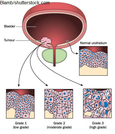
Are the cells well differentiated? Meaning they have an appearance and are arranged in a way that is similar to normal cells. If this is the case, these types of cancer tend to grow and spread slowly.
Or are they hard to differentiate (moderately or poorly differentiated)? Meaning their appearance and arrangement doesn’t mirror what normal healthy cells in that particular area possess, and they look abnormal. These types tend to spread and grow quickly.
The tumor grades of low to high can be given along with numerical values to reflect that rating. However, it’s important to note that some forms of cancer have they own grading systems, such as breast cancer which uses the Nottingham Score System.
Numerical values for tumor grading include: Grade I to either Grade III or IV
Here’s an example:
Grade I: well differentiated, low grade
Grade II: moderately differentiated, intermediate grade…cells have some differentiation
Grade III: poorly differentiated, high grade
Grade IV: undifferentiated, high grade…cells are extremely abnormal
Here you can see an illustration of this based on Grade I to III.
Tumor Staging:
Staging tells about the main tumor (location, size etc.), and if the cancer has spread to other parts of the body like the lymph nodes or other organs.
This is useful information because it will help with:
- Developing a treatment plan….treatment for an earlier stage of cancer is different than a later stage
- Potential clinical trial participation
- Providing some insight on how aggressive and treatable the cancer may be
The patient will need tests to help determine the stage: MRI, CT SCAN, ultrasound, blood work, physical assessment findings etc.
It’s important to note that the cancer stage designated at the time of the cancer diagnosis doesn’t change, and if the cancer spreads or metastases that information is added onto the original category designation. In addition, if the cancer is restaged at some point, the restage doesn’t replace the original stage designation but is included with it.
There are different types of staging systems used to stage cancer, and this is determined by what type of cancer the patient has.
One system you want to be familiar with as the nurse is the TNM staging system. This system is used to stage cancers with solid tumors like that found in colon cancer and other types. Therefore, it’s not used for blood cancers, brain, spinal cord.
TNM is an acronym that is made up of categories that stands for: Tumor, Nodal Involvement, Metastasis
Tumor (primary): this category details the location and how much of that tumor is growing into other tissues (is it just hanging out by itself in its primary site or is it growing inside other structures and layers)…the higher the number the more it has grown into other layers or structures
TX: tumor can’t be measured
T0: no tumor is found
Tis: tumor is in situ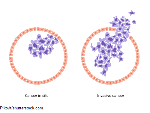
- In situ means: in original place….the tumor is found in its original place and has not spread from its original location
- Not cancerous at this time but in the future it could turn cancerous and spread
T1, T2, T3, T4: describes the size/amount the tumor has grown and affected other areas
- Higher the number the larger the size/amount it has grown into other areas….example: T1 is smaller than a T4
- Depending on the cancer type sometimes letters can be added after the number to further describe the tumor’s growth
- For example, a breast cancer tumor can be described as T1a: tumor is greater than 1 mm but less than 5 mm or T4a: tumor has spread to the chest wall
Nodes (regional lymph node): this category details the spread of the cancer in a nearby lymph node (closest to the primary tumor).
- Unfortunately, parts of the tumor can break off and collect in the lymphatic system, hence nearby lymph nodes. Lymph nodes are small clustered structures that help us fight infection. They are found around many important organs.
NX: cancer in regional lymph node can’t be measured
N0: no cancer present in regional lymph node
N1, N2, N3: the number and location of the lymph nodes that have cancer. N3 means there are more lymph nodes that contain cancer than N1.
- Like with T (tumor), letters can be added after the number to further describe the spread of the cancer to the lymph nodes. Example N2a or N2b etc.
Metastasis: details if the cancer (primary tumor) has spread to other parts of the body, and if this is the case, how much and the location of it.
M0: no cancer found in other parts
M1: cancer has spread to organs and tissues
- Again, depending on the cancer, letters can be added after the number to further describe the metastasis of the cancer to other parts of the body.
Other Add-Ons with the TNM system: As the nurse, you may see letters added to the TNM letters that provide further information the category.
A lowercase “c” or “p” can be in front of a category of the TNM classification. (Example: cT1 or pN2)
- A lowercase “c” means clinical staging. This means that the TNM classification was given before treatment began and is based on test results and assessment findings.
- A lowercase “p” means pathological staging. This means the staging classification was given based on the findings after surgery. It takes into account the test results and assessment findings of the clinical stage, but it can give a more detailed picture of the cancer. For example, the surgery may find that the cancer has metastasized further throughout the body than previously seen with test results. However, not all patients have surgery and in this case a pathological stage can’t be determined.
A lowercase “y” or “r” can be in front a TNM category example: ycT3 or rpT3
y: This represents post-therapy staging. This is given after therapy was administered (example: chemo) and how much of the tumor remains before surgery.
A lowercase “r” is to show reoccurrence of the cancer.
The TNM system is used to calculate the number system: Stage 0, I, II, II, IV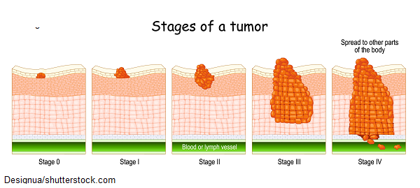
Stage 0: cancer in situ….the cancer is still in its original place…hasn’t invaded surrounding tissues
Stage I: the cancer is localized and not spread into other tissues or lymph nodes
Stage II: spread into surrounding tissues and nearby lymph nodes
Stage III: has spread to even deeper tissues than stage II and further away lymph nodes BUT has not spread to other distant structures in the body like organs
Stage IV: most advanced…called metastatic cancer…it’s cancer that has spread to other parts of the body beyond where the cancer started
References:
Cancer Staging. National Cancer Institute. (2022). Retrieved 7 January 2022, from https://www.cancer.gov/about-cancer/diagnosis-staging/staging.