This article will explain how to assess the chest (heart and lungs) as a nurse. This assessment is part of the nursing head-to-toe assessment you have to perform in nursing school and on the job.
During the chest assessment you will be assessing the following structures:
- Overall appearance of the chest
- Lung Sounds: includes abnormal lung sounds
- Heart Sounds
Video Demonstration on Nursing Chest Assessment
Chest:
Inspect the chest
- Is the respiratory effort easy? Is the patient using the abdominal or accessory muscles for breathing?
- Does the patient have a barreled chest (some patients with COPD do)?
- Assess the skin for wounds, pacemaker present, subcutaneous port etc.?
Heart Sounds:
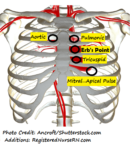
Auscultate heart sounds at 5 locations, specifically valve locations:
- Remember the mnemonic: “All Patients Effectively (Erb’s Point…halfway point between the base and apex of the heart) Take Medicine”
- All: Aortic
- Patients: Pulmonic
- Effectively: Erb’s Point (no valve at this location)
- Take: Tricuspid
- Medicine: Mitral
- Use diaphragm of stethoscope: listening for lub dub (S1 and S2…any splits) and the rhythm: is it regular (if on cardiac monitor…note heart rhythm)
Aortic: found right of the sternal border in the 2nd intercostal space REPRESENTS S2 “dub” which is the loudest.
Pulmonic: found left of the sternal border in the 2nd intercostal space REPRESENTS S2 “dub” which is the loudest.
Erb’s Point: found left of the sternal border in the 3rd intercostal space…no valve here just the halfway point.
Tricuspid: found left of the sternal border in the 4th intercostal space REPRESENTS S1 “lub”.
Mitral: found midclavicular in the 5th intercostal space REPRESENTS S1 “lub” (also the site of point of maximal impulse) APICAL PULSE….count pulse for 1 full minute.
Then listen with the BELL of the stethoscope at the same locations: for a blowing or swooshing noise…heart murmur.
Lung Sounds:
If you would like to hear some abnormal lung sounds, please watch our video called “abnormal lung sounds”.
Auscultate anteriorly:
- Start at: the apex of the lung which is right above the clavicle
- Then move to the 2nd intercostal space to assess the right and left upper lobes.
- Move to the 4th intercostal space, you will be assessing the right middle lobe and the left upper lobe.
- Lastly move to the mid-axillary are at the 6th intercostal space and you will be assessing the right and left lower lobes.

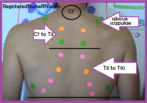
Auscultate posteriorly:
- Start right above the scapulae to listen to the apex of the lungs.
- Then find C7 (which is the vertebral prominence) and go to T3…in between the shoulder blades and spine. This will assess the right and left upper lobes.
- Then from T3 to T10 you will be able to assess the right and left lower lobes.
You may be interested in watching a complete head-to-toe assessment.